
Search Count: 53
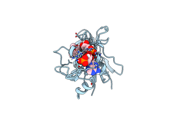 |
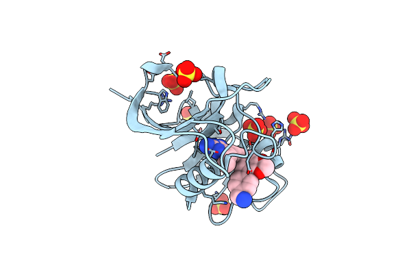
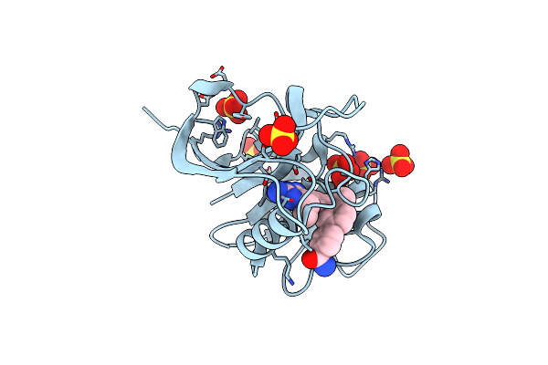
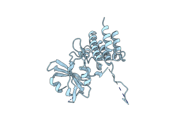
Crystal Structure Of Mycobacterium Avium Dihydrofolate Reductase In Complex With Nadph And Trimethoprim
Organism: Mycobacterium avium
Method: X-RAY DIFFRACTION Resolution:2.00 Å Release Date: 2025-03-19 Classification: OXIDOREDUCTASE/INHIBITOR Ligands: NDP, TOP, CL |
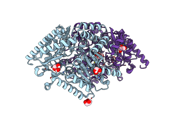 |
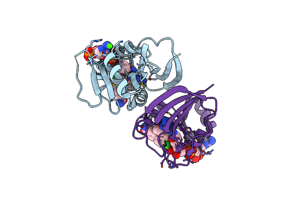
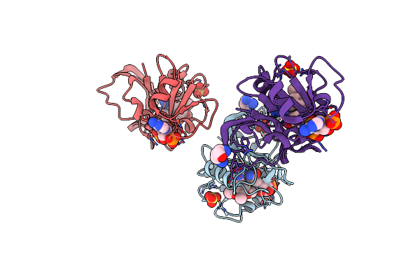
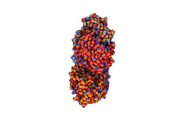
Crystal Structure Of The Dimeric Transaminase Doed From C. Salexigens Dsm 3043.
Organism: Chromohalobacter israelensis dsm 3043
Method: X-RAY DIFFRACTION Release Date: 2025-03-12 Classification: TRANSFERASE Ligands: GOL, PLP |
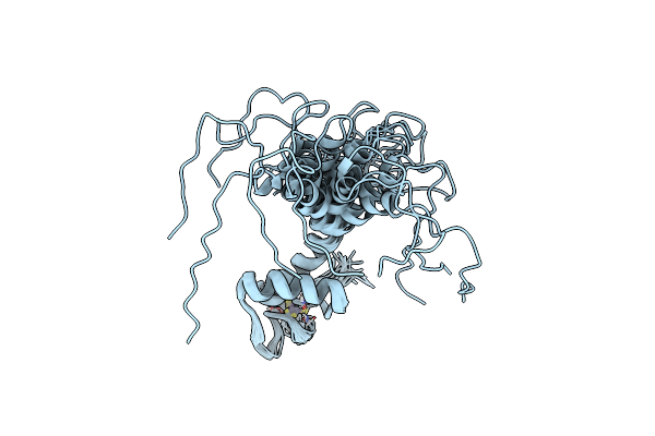 |
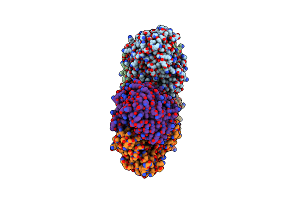
Organism: Homo sapiens
Method: SOLUTION NMR Release Date: 2023-10-18 Classification: LIGASE Ligands: ZN |
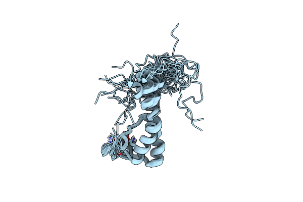 |
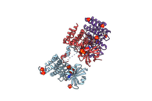
Organism: Homo sapiens
Method: SOLUTION NMR Release Date: 2023-07-05 Classification: LIGASE Ligands: ZN |
 |
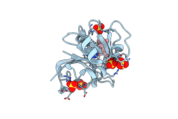
E. Coli Dihydrofolate Reductase Complexed With 5-(3-(7-(4-(Aminomethyl)Phenyl)Benzo[D][1,3]Dioxol-5-Yl)But-1-Yn-1-Yl)-6-Ethylpyrimidine-2,4-Diamine (Ucp1223)
Organism: Escherichia coli
Method: X-RAY DIFFRACTION Resolution:1.91 Å Release Date: 2022-07-27 Classification: OXIDOREDUCTASE Ligands: SO4, 560 |
 |
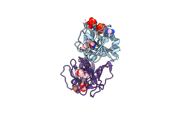
Dfra1 Complexed With Nadph And 4'-Chloro-3'-(4-(2,4-Diamino-6-Ethylpyrimidin-5-Yl)But-3-Yn-2-Yl)-[1,1'-Biphenyl]-4-Carboxamide (Ucp1228)
Organism: Escherichia coli
Method: X-RAY DIFFRACTION Resolution:1.77 Å Release Date: 2022-07-27 Classification: OXIDOREDUCTASE Ligands: 4SI, NAP, CA |
 |
Crystal Structure Of Escherichia Coli Dihydrofolate Reductase In Complex With Trimethoprim
Organism: Escherichia coli (strain k12)
Method: X-RAY DIFFRACTION Resolution:2.35 Å Release Date: 2022-06-29 Classification: OXIDOREDUCTASE Ligands: TOP, SO4 |
 |
Crystal Structure Of Dfra5 Dihydrofolate Reductase In Complex With Trimethoprim And Nadph
Organism: Escherichia coli
Method: X-RAY DIFFRACTION Resolution:2.61 Å Release Date: 2022-06-29 Classification: OXIDOREDUCTASE Ligands: TOP, NDP, SO4 |
 |
Crystal Structure Of Dfra1 Dihydrofolate Reductase In Complex With Trimethoprim
Organism: Klebsiella pneumoniae
Method: X-RAY DIFFRACTION Resolution:2.15 Å Release Date: 2022-06-01 Classification: OXIDOREDUCTASE Ligands: TOP |
 |
Crystal Structure Of Escherichia Coli Dihydrofolate Reductase In Complex With Trimethoprim And Nadph
Organism: Escherichia coli
Method: X-RAY DIFFRACTION Resolution:3.04 Å Release Date: 2022-06-01 Classification: OXIDOREDUCTASE Ligands: ARG, NAP, TOP, SO4 |
 |
Dfra1 Complexed With Nadph And 5-(3-(7-(4-(Aminomethyl)Phenyl)Benzo[D][1,3]Dioxol-5-Yl)But-1-Yn-1-Yl)-6-Ethylpyrimidine-2,4-Diamine (Ucp1223)
Organism: Escherichia coli
Method: X-RAY DIFFRACTION Resolution:1.44 Å Release Date: 2022-05-25 Classification: OXIDOREDUCTASE/OXIDOREDUCTASE INHIBITOR Ligands: NAP, CA, 8MU, 560 |
 |
Dfra5 Complexed With Nadph And 5-(3-(7-(4-(Aminomethyl)Phenyl)Benzo[D][1,3]Dioxol-5-Yl)But-1-Yn-1-Yl)-6-Ethylpyrimidine-2,4-Diamine (Ucp1223)
Organism: Escherichia coli
Method: X-RAY DIFFRACTION Resolution:2.19 Å Release Date: 2022-05-25 Classification: OXIDOREDUCTASE/OXIDOREDUCTASE INHIBITOR Ligands: NAP, SO4, 560 |
 |
Dfra5 Complexed With Nadph And 4'-Chloro-3'-(4-(2,4-Diamino-6-Ethylpyrimidin-5-Yl)But-3-Yn-2-Yl)-[1,1'-Biphenyl]-4-Carboxamide (Ucp1228)
Organism: Escherichia coli
Method: X-RAY DIFFRACTION Resolution:1.92 Å Release Date: 2022-05-25 Classification: OXIDOREDUCTASE/OXIDOREDUCTASE INHIBITOR Ligands: NAP, GOL, 4SI, SO4 |
 |
E. Coli Dihydrofolate Reductase Complexed With 4'-Chloro-3'-(4-(2,4-Diamino-6-Ethylpyrimidin-5-Yl)But-3-Yn-2-Yl)-[1,1'-Biphenyl]-4-Carboxamide (Ucp1228)
Organism: Escherichia coli
Method: X-RAY DIFFRACTION Resolution:2.10 Å Release Date: 2022-05-11 Classification: OXIDOREDUCTASE Ligands: ZM4, SO4 |
 |
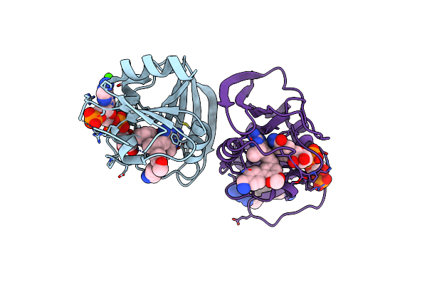
Crystal Structure Of Escherichia Coli Enolase Complexed With A Natural Inhibitor Sf2312.
Organism: Escherichia coli
Method: X-RAY DIFFRACTION Resolution:2.24 Å Release Date: 2019-11-27 Classification: LYASE/LYASE inhibitor Ligands: 4NG, MG, GOL, SO4 |
 |
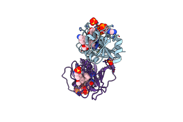
Structure Of E.Coli Enolase In Complex With An Analog Of The Natural Product Sf-2312 Metabolite.
Organism: Escherichia coli
Method: X-RAY DIFFRACTION Resolution:2.57 Å Release Date: 2019-11-27 Classification: LYASE Ligands: TLA, SO4, MG, GOL, KVM |
 |
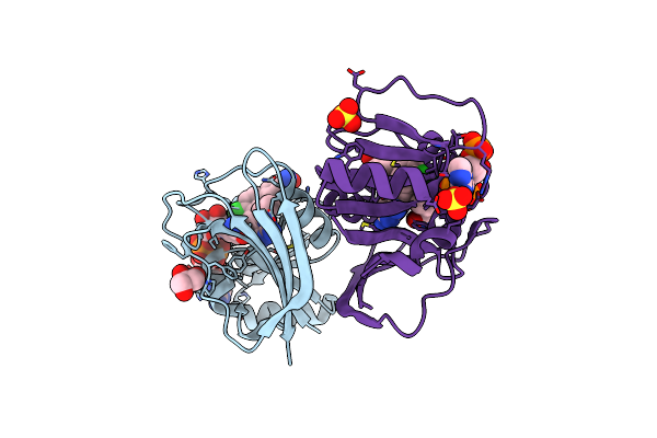
Crystal Structure Of Unphosphorylated Human Pkr Kinase Domain In Complex With Adp
Organism: Homo sapiens
Method: X-RAY DIFFRACTION Resolution:2.60 Å Release Date: 2019-07-10 Classification: TRANSFERASE Ligands: ADP, PO4, MG, SO4 |
 |
Organism: Homo sapiens
Method: X-RAY DIFFRACTION Resolution:3.10 Å Release Date: 2019-07-10 Classification: TRANSFERASE |
 |
Crystal Structure Of Mutant B1 Immunoglobulin-Binding Domain Of Streptococcal Protein G (T16F, T18A, V21E, T25L, K28Y, V29I, K31R, Q32H, Y33L, N35K, D36H, N37Q)
Organism: Streptococcus
Method: X-RAY DIFFRACTION Resolution:1.40 Å Release Date: 2019-01-23 Classification: DE NOVO PROTEIN Ligands: ZN, CL |
 |
Crystal Structure Of B1 Immunoglobulin-Binding Domain Of Streptococcal Protein G (T16F, T18A, V21H, T25H, K28Y, V29I, K31R, Q32A, Y33L, N35K, D36A, N37Q)
Organism: Streptococcus
Method: X-RAY DIFFRACTION Resolution:1.40 Å Release Date: 2019-01-23 Classification: METAL BINDING PROTEIN Ligands: ZN, ACT, NA, CL, PO4, DPO |