
Search Count: 112
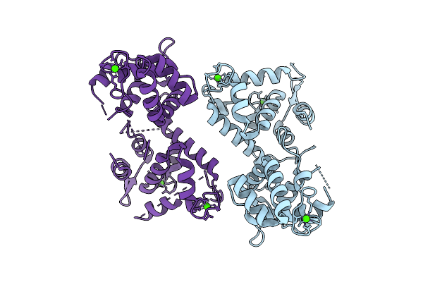 |
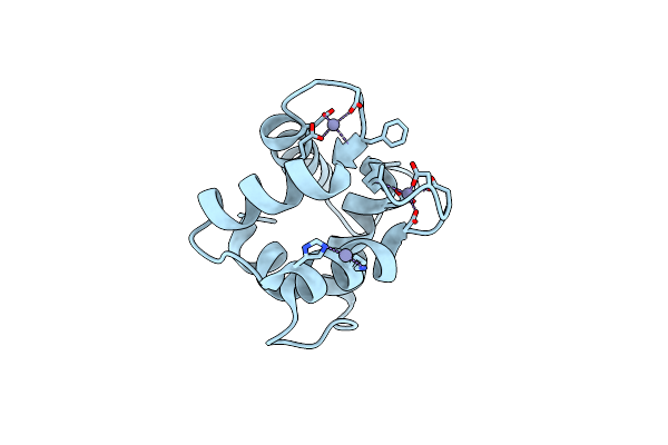
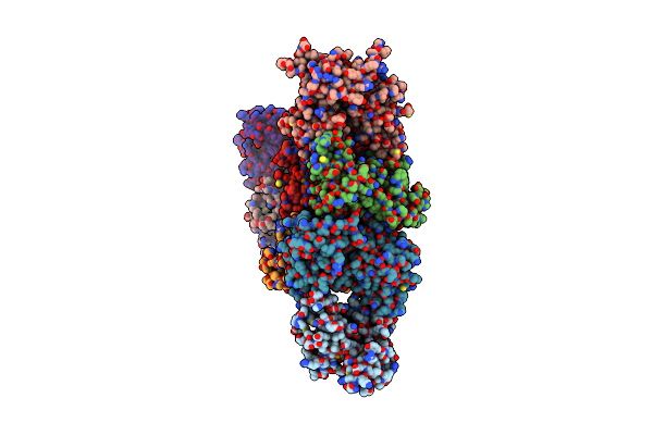
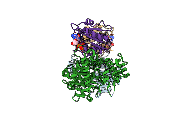
Crystal Structure Of The Dictyostelium Discoideum Mitochondrial Calcium Uptake Protein (Ddmicu)
Organism: Dictyostelium discoideum
Method: X-RAY DIFFRACTION Release Date: 2025-07-30 Classification: METAL BINDING PROTEIN Ligands: CA |
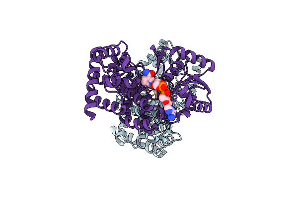 |
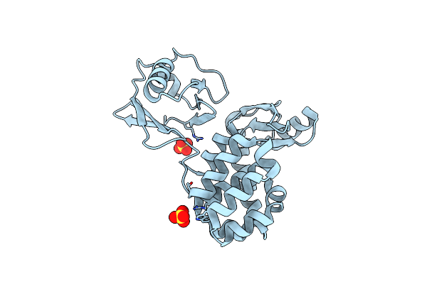
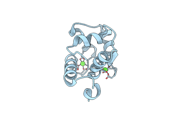
Crystal Structure Of Glycerol Dehydrogenase In The Presence Of Nad+ And Glycerol
Organism: Escherichia coli k-12
Method: X-RAY DIFFRACTION Resolution:2.00 Å Release Date: 2024-03-27 Classification: OXIDOREDUCTASE Ligands: ZN, NAD, GOL |
 |
Organism: Escherichia coli k-12
Method: X-RAY DIFFRACTION Resolution:2.90 Å Release Date: 2023-06-14 Classification: OXIDOREDUCTASE Ligands: ZN, TRS |
 |
Organism: Escherichia coli k-12
Method: X-RAY DIFFRACTION Resolution:2.60 Å Release Date: 2023-06-14 Classification: OXIDOREDUCTASE Ligands: ZN, NAD, TRS |
 |
Organism: Mus musculus
Method: X-RAY DIFFRACTION Resolution:2.80 Å Release Date: 2023-03-15 Classification: METAL BINDING PROTEIN Ligands: CA, GOL, ZN |
 |
Organism: Mus musculus
Method: X-RAY DIFFRACTION Resolution:1.72 Å Release Date: 2023-03-15 Classification: METAL BINDING PROTEIN Ligands: ZN, GOL |
 |
Organism: Homo sapiens
Method: X-RAY DIFFRACTION Resolution:2.60 Å Release Date: 2023-03-15 Classification: METAL BINDING PROTEIN Ligands: ZN |
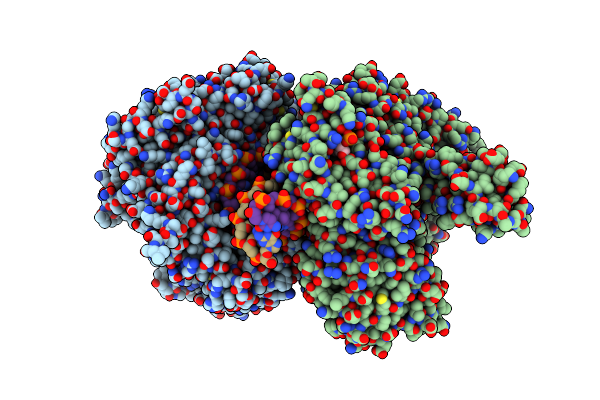 |
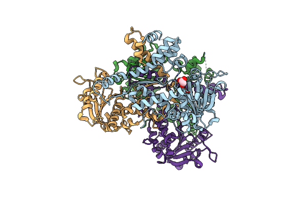
Crystal Structure Of Replisomal Dimer Of Dna Polymerase From Bacteriophage Rb69 With Dna Duplexes
Organism: Enterobacteria phage rb69, Synthetic construct
Method: X-RAY DIFFRACTION Resolution:2.20 Å Release Date: 2021-07-14 Classification: TRANSFERASE/DNA Ligands: DUP, CA, 5GP, MG |
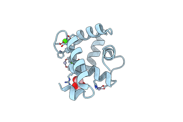 |
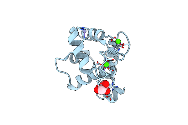
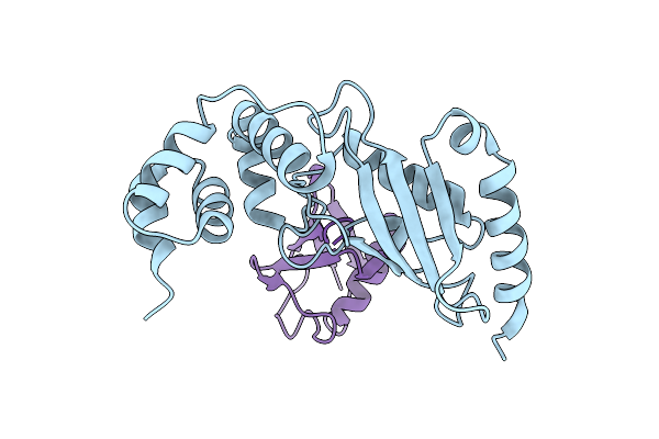
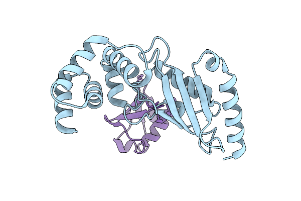
Crystal Structure Of The Efhd1/Swiprosin-2, A Mitochondrial Actin-Binding Protein
Organism: Mus musculus
Method: X-RAY DIFFRACTION Resolution:2.07 Å Release Date: 2021-01-06 Classification: METAL BINDING PROTEIN Ligands: CA, ZN, GOL |
 |
Crystal Structure Of The N-Terminal Domain Single Mutant (D119A) Of The Human Mitochondrial Calcium Uniporter Fused With T4 Lysozyme
Organism: Enterobacteria phage t4, Homo sapiens
Method: X-RAY DIFFRACTION Resolution:2.85 Å Release Date: 2020-05-27 Classification: TRANSPORT PROTEIN Ligands: SO4 |
 |
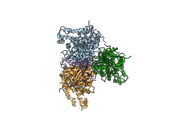
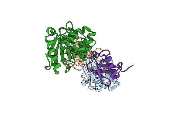
Crystal Structure Of The Mitochondrial Calcium Uptake 1 And 2 Heterodimer (Micu1-Micu2 Heterodimer) In An Apo State
Organism: Homo sapiens
Method: X-RAY DIFFRACTION Resolution:3.10 Å Release Date: 2020-03-04 Classification: METAL BINDING PROTEIN |
 |
Crystal Structure Of The N-Terminal Domain Single Mutant (S92E) Of The Human Mitochondrial Calcium Uniporter Fused With T4 Lysozyme
Organism: Enterobacteria phage t4, Homo sapiens
Method: X-RAY DIFFRACTION Resolution:2.50 Å Release Date: 2020-02-19 Classification: TRANSPORT PROTEIN Ligands: SO4 |
 |
Organism: Vibrio vulnificus mo6-24/o
Method: X-RAY DIFFRACTION Resolution:2.62 Å Release Date: 2019-08-21 Classification: TOXIN |
 |
Crystal Structure Of The Makes Caterpillars Floppy (Mcf)-Like Effector Of Vibrio Vulnificus Mo6-24/O
Organism: Vibrio vulnificus
Method: X-RAY DIFFRACTION Resolution:2.36 Å Release Date: 2019-08-07 Classification: TOXIN Ligands: GOL |
 |
Crystal Structure Of Alpha-Beta Hydrolase (Abh) And Makes Caterpillars Floppy (Mcf)-Like Effectors Of Vibrio Vulnificus Mo6-24/O
Organism: Vibrio vulnificus
Method: X-RAY DIFFRACTION Resolution:3.50 Å Release Date: 2019-08-07 Classification: TOXIN |
 |
Crystal Structure Of The Makes Caterpillars Floppy (Mcf)-Like Effector Of Vibrio Vulnificus Mo6-24/O In Complex With A Human Adp-Ribosylation Factor 3 (Arf3)
Organism: Vibrio vulnificus, Homo sapiens
Method: X-RAY DIFFRACTION Resolution:2.10 Å Release Date: 2019-08-07 Classification: TOXIN/PROTEIN BINDING Ligands: GTP, MG |
 |
Crystal Structure Of Ub-Conjugated Ube2K C92K&K97A Mutant (Isopeptide Linkage), 2.7 A Resolution
Organism: Homo sapiens
Method: X-RAY DIFFRACTION Resolution:2.70 Å Release Date: 2019-03-20 Classification: LIGASE |
 |
Crystal Structure Of Ub-Conjugated Ube2K C92K&K97A Mutant (Isopeptide Linkage), 2.1 A Resolution
Organism: Homo sapiens
Method: X-RAY DIFFRACTION Resolution:2.10 Å Release Date: 2019-03-20 Classification: LIGASE |
 |
Organism: Homo sapiens
Method: X-RAY DIFFRACTION Resolution:2.47 Å Release Date: 2018-11-21 Classification: LIGASE |
 |
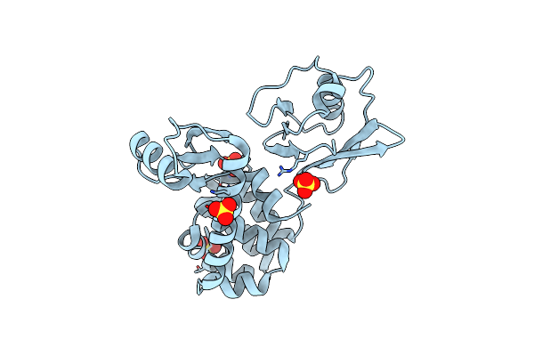
Organism: Homo sapiens
Method: X-RAY DIFFRACTION Resolution:1.86 Å Release Date: 2017-09-13 Classification: METAL BINDING PROTEIN Ligands: CA |