
Search Count: 18
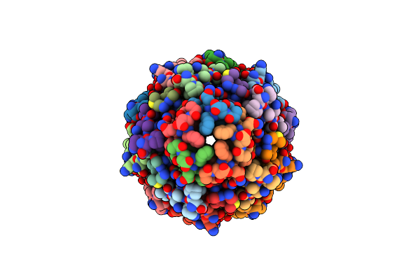 |
Organism: Enterobacteria phage m13
Method: ELECTRON MICROSCOPY Release Date: 2024-01-03 Classification: VIRAL PROTEIN |
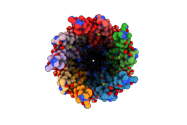 |
Organism: Enterobacteria phage m13
Method: ELECTRON MICROSCOPY Release Date: 2024-01-03 Classification: VIRAL PROTEIN |
 |
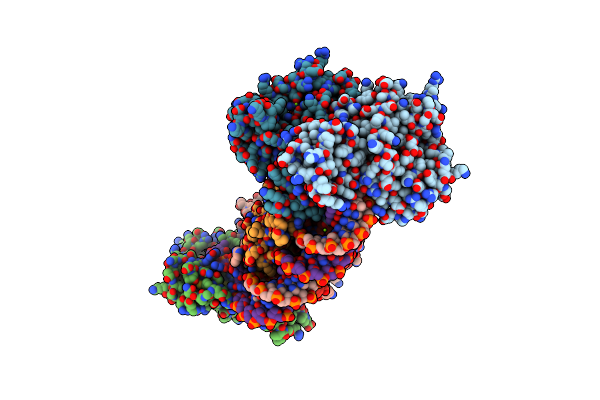
Organism: Enterobacteria phage m13
Method: ELECTRON MICROSCOPY Release Date: 2023-08-16 Classification: VIRAL PROTEIN |
 |
Organism: Enterobacteria phage m13
Method: SOLUTION NMR Release Date: 2019-08-28 Classification: DE NOVO PROTEIN |
 |
Organism: Enterobacteria phage m13
Method: SOLUTION NMR Release Date: 2019-08-28 Classification: DE NOVO PROTEIN |
 |
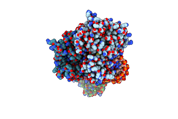
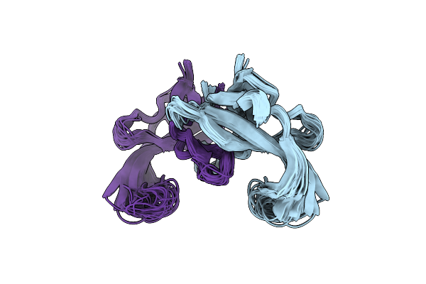
Organism: Homo sapiens, Enterobacteria phage m13
Method: X-RAY DIFFRACTION Resolution:2.00 Å Release Date: 2019-07-24 Classification: LIGASE |
 |
Crystal Structure The Escherichia Coli Cas1-Cas2 Complex Bound To Protospacer Dna
Organism: Escherichia coli (strain k12), Enterobacteria phage m13
Method: X-RAY DIFFRACTION Resolution:3.20 Å Release Date: 2015-10-28 Classification: Hydrolase/DNA |
 |
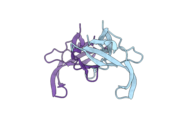
Crystal Structure The Escherichia Coli Cas1-Cas2 Complex Bound To Protospacer Dna And Mg
Organism: Escherichia coli (strain k12), Enterobacteria phage m13
Method: X-RAY DIFFRACTION Resolution:2.95 Å Release Date: 2015-10-28 Classification: Hydrolase/DNA Ligands: MG |
 |
Crystal Structure The Escherichia Coli Cas1-Cas2 Complex Bound To Protospacer Dna With Splayed Ends
Organism: Escherichia coli (strain k12), Enterobacteria phage m13
Method: X-RAY DIFFRACTION Resolution:3.35 Å Release Date: 2015-10-28 Classification: Hydrolase/DNA |
 |
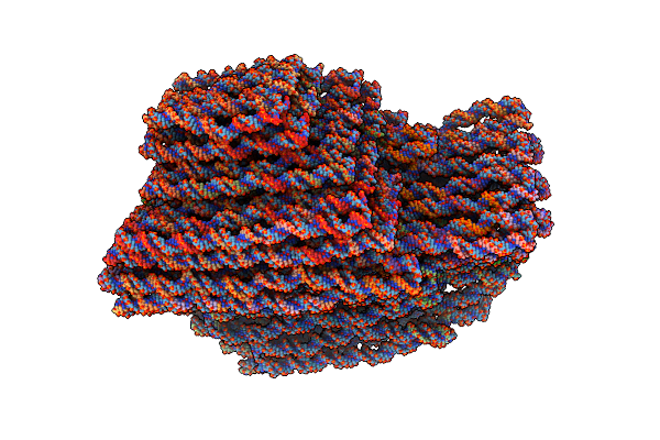
Capsid Model Of M13 Bacteriophage Virus From Magic-Angle Spinning Nmr And Rosetta Modeling
Organism: Enterobacteria phage m13
Method: SOLID-STATE NMR Release Date: 2015-01-07 Classification: VIRAL PROTEIN |
 |
Organism: Enterobacteria phage m13, Synthetic construct
Method: ELECTRON MICROSCOPY Resolution:11.50 Å Release Date: 2014-07-09 Classification: DNA |
 |
Fusion Of N-Terminal Domain Of The Minor Coat Protein From Gene Iii In Phage M13, And C-Terminal Domain Of E. Coli Protein-Tola
Organism: Enterobacteria phage m13, Escherichia coli
Method: X-RAY DIFFRACTION Resolution:1.85 Å Release Date: 1999-05-20 Classification: VIRAL PROTEIN |
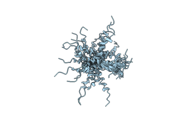 |
Solution Nmr Structures Of The Major Coat Protein Of Filamentous Bacteriophage M13 Solubilized In Dodecylphosphocholine Micelles, 25 Lowest Energy Structures
Organism: Enterobacteria phage m13
Method: SOLUTION NMR Release Date: 1998-11-11 Classification: VIRAL PROTEIN |
 |
Solution Nmr Structures Of The Major Coat Protein Of Filamentous Bacteriophage M13 Solubilized In Sodium Dodecyl Sulphate Micelles, 25 Lowest Energy Structures
Organism: Enterobacteria phage m13
Method: SOLUTION NMR Release Date: 1998-11-11 Classification: VIRAL PROTEIN |
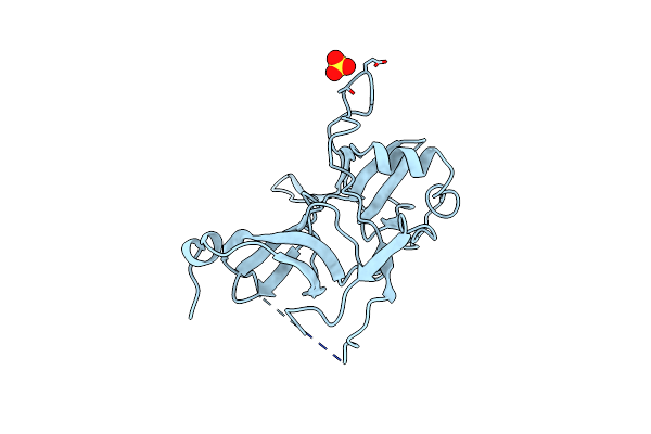 |
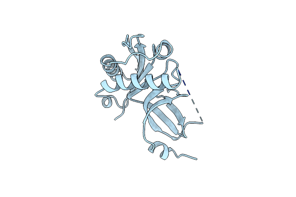
Crystal Structure Of The N-Terminal Domains Of Bacteriophage Minor Coat Protein G3P
Organism: Enterobacteria phage m13
Method: X-RAY DIFFRACTION Resolution:1.46 Å Release Date: 1998-01-28 Classification: VIRAL PROTEIN Ligands: SO4 |
 |
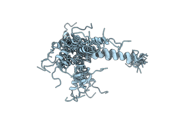
Refined Solution Structure Of The Tyr 41--> His Mutant Of The M13 Gene V Protein. A Comparison With The Crystal Structure
Organism: Enterobacteria phage m13
Method: SOLUTION NMR Release Date: 1995-10-15 Classification: DNA-BINDING (VIRAL) |
 |
Refined Solution Structure Of The Tyr 41--> His Mutant Of The M13 Gene V Protein. A Comparison With The Crystal Structure
Organism: Enterobacteria phage m13
Method: SOLUTION NMR Release Date: 1995-10-15 Classification: DNA-BINDING (VIRAL) |
 |
Organism: Enterobacteria phage m13
Method: X-RAY DIFFRACTION Resolution:2.30 Å Release Date: 1986-01-21 Classification: DNA BINDING (VIRAL) |