
Search Count: 39
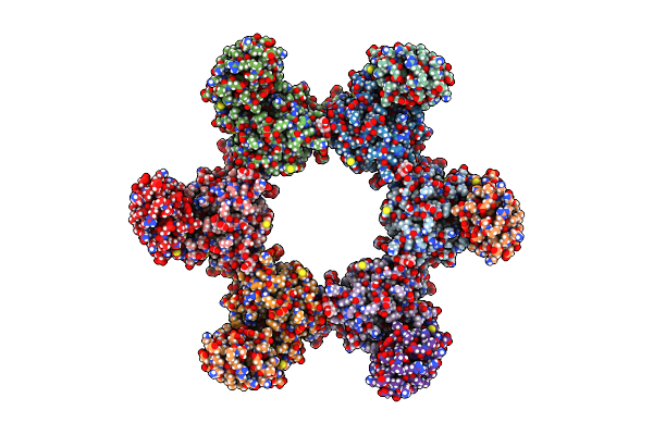 |
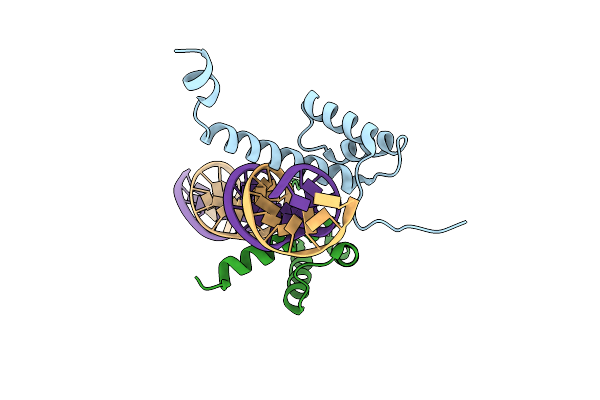
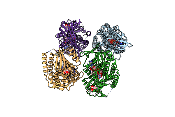
Cryo-Em Structure Of Cowpox Virus M2 In Complex With Human B7.1 (Hexameric Ring)
Organism: Cowpox virus (brighton red), Homo sapiens
Method: ELECTRON MICROSCOPY Release Date: 2025-02-05 Classification: VIRAL PROTEIN Ligands: NAG |
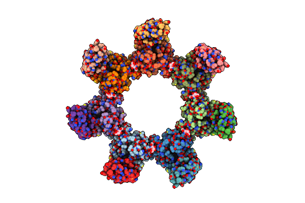 |
Cryo-Em Structure Of Cowpox Virus M2 In Complex With Human B7.2 (Heptameric Ring)
Organism: Cowpox virus (brighton red), Homo sapiens
Method: ELECTRON MICROSCOPY Release Date: 2025-02-05 Classification: VIRAL PROTEIN Ligands: NAG |
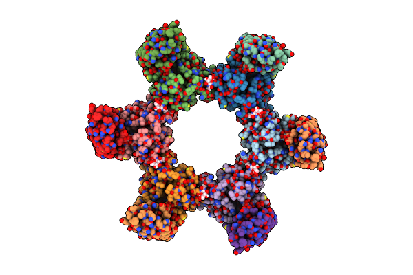 |
Cryo-Em Structure Of Cowpox Virus M2 In Complex With Human B7.2 (Hexameric Ring)
Organism: Cowpox virus (brighton red), Homo sapiens
Method: ELECTRON MICROSCOPY Release Date: 2025-02-05 Classification: VIRAL PROTEIN Ligands: NAG |
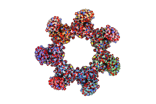 |
Cryo-Em Structure Of Cowpox Virus M2 In Complex With Human B7.1 (Heptameric Ring)
Organism: Cowpox virus (brighton red), Homo sapiens
Method: ELECTRON MICROSCOPY Release Date: 2025-02-05 Classification: VIRAL PROTEIN Ligands: NAG |
 |
Crystal Structure Of Human Two Pore Domain Potassium Ion Channel Trek-2 (K2P10.1) In Complex With A Nanobody (Nb58)
Organism: Homo sapiens, Lama glama
Method: X-RAY DIFFRACTION Resolution:3.59 Å Release Date: 2024-05-29 Classification: MEMBRANE PROTEIN Ligands: K |
 |
Crystal Structure Of Human Two Pore Domain Potassium Ion Channel Trek-2 (K2P10.1) In Complex With An Inhibitory Nanobody (Nb61)
Organism: Homo sapiens, Lama glama
Method: X-RAY DIFFRACTION Resolution:3.50 Å Release Date: 2024-05-29 Classification: MEMBRANE PROTEIN Ligands: K |
 |
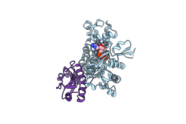
Crystal Structure Of Human Two Pore Domain Potassium Ion Channel Trek-2 (K2P10.1) In Complex With An Activatory Nanobody (Nb67)
Organism: Homo sapiens, Lama glama
Method: X-RAY DIFFRACTION Resolution:2.40 Å Release Date: 2024-05-29 Classification: MEMBRANE PROTEIN Ligands: K, MPD |
 |
Crystal Structure Of Human Two Pore Domain Potassium Ion Channel Trek-2 (K2P10.1) In Complex With An Activatory Nanobody (Nb76)
Organism: Homo sapiens, Lama glama
Method: X-RAY DIFFRACTION Resolution:3.20 Å Release Date: 2024-05-29 Classification: MEMBRANE PROTEIN Ligands: BA, Y01 |
 |
Organism: Medicago truncatula
Method: X-RAY DIFFRACTION Resolution:2.10 Å Release Date: 2021-08-11 Classification: PLANT PROTEIN/DNA |
 |
Organism: Mus musculus, Oryctolagus cuniculus
Method: X-RAY DIFFRACTION Resolution:2.56 Å Release Date: 2020-12-09 Classification: PROTEIN BINDING Ligands: ATP, CA |
 |
Human Plasmakallikrein Protease Domain In Complex With Active Site Directed Inhibitor
Organism: Homo sapiens
Method: X-RAY DIFFRACTION Resolution:1.42 Å Release Date: 2020-07-08 Classification: HYDROLASE Ligands: GSH, DMS, PO4, GOL, MU8 |
 |
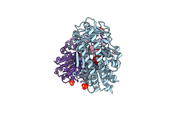
Coagulation Factor Xi Protease Domain In Complex With Active Site Inhibitor
Organism: Homo sapiens
Method: X-RAY DIFFRACTION Resolution:1.17 Å Release Date: 2020-07-08 Classification: HYDROLASE Ligands: SO4, DMS, NA, J7B |
 |
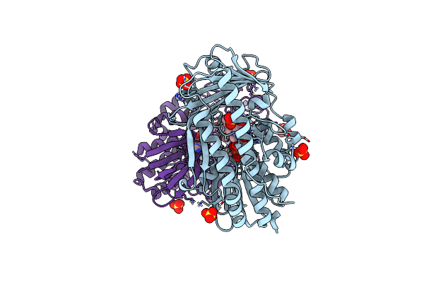
Coagulation Factor Xi Protease Domain In Complex With Active Site Inhibitor
Organism: Homo sapiens
Method: X-RAY DIFFRACTION Resolution:1.29 Å Release Date: 2020-07-08 Classification: HYDROLASE Ligands: NW2, SO4, DMS |
 |
Coagulation Factor Xi Protease Domain In Complex With Active Site Inhibitor
Organism: Homo sapiens
Method: X-RAY DIFFRACTION Resolution:1.33 Å Release Date: 2020-07-08 Classification: HYDROLASE Ligands: SO4, DMS, NW5 |
 |
Coagulation Factor Xi Protease Domain In Complex With Active Site Inhibitor
Organism: Homo sapiens
Method: X-RAY DIFFRACTION Resolution:2.63 Å Release Date: 2020-07-08 Classification: HYDROLASE Ligands: NWE |
 |
Organism: Homo sapiens
Method: X-RAY DIFFRACTION Resolution:1.26 Å Release Date: 2020-07-01 Classification: HYDROLASE Ligands: QGS |
 |
Organism: Uncultured bacterium
Method: X-RAY DIFFRACTION Resolution:2.10 Å Release Date: 2017-05-10 Classification: OXIDOREDUCTASE Ligands: SO4, FAD |
 |
Crystal Structure Of Tetracycline Destructase Tet(50) In Complex With Anhydrotetracycline
Organism: Uncultured bacterium
Method: X-RAY DIFFRACTION Resolution:2.25 Å Release Date: 2017-05-10 Classification: OXIDOREDUCTASE Ligands: SO4, FAD, TDC |
 |
Crystal Structure Of Tetracycline Destructase Tet(50) In Complex With Chlortetracycline
Organism: Uncultured bacterium
Method: X-RAY DIFFRACTION Resolution:1.75 Å Release Date: 2017-05-10 Classification: OXIDOREDUCTASE Ligands: SO4, FAD, CTC |
 |
Organism: Uncultured bacterium
Method: X-RAY DIFFRACTION Resolution:1.85 Å Release Date: 2017-05-10 Classification: OXIDOREDUCTASE Ligands: GOL, FAD |