
Search Count: 24
 |
Organism: Saccharomyces cerevisiae
Method: ELECTRON MICROSCOPY Release Date: 2025-05-14 Classification: PROTEIN TRANSPORT |
 |
Organism: Saccharomyces cerevisiae
Method: ELECTRON MICROSCOPY Release Date: 2025-04-30 Classification: PROTEIN TRANSPORT |
 |
Organism: Chlamydomonas reinhardtii
Method: ELECTRON MICROSCOPY Release Date: 2025-03-05 Classification: PHOTOSYNTHESIS |
 |
Organism: Listeria monocytogenes 10403s
Method: ELECTRON MICROSCOPY Release Date: 2024-06-19 Classification: TOXIN |
 |
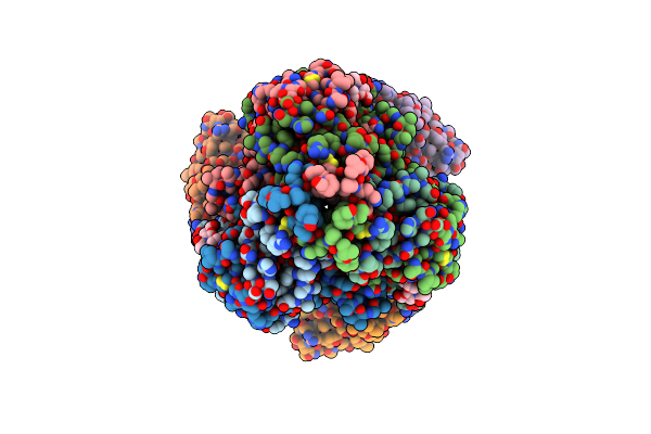
Organism: Homo sapiens
Method: ELECTRON MICROSCOPY Release Date: 2024-03-27 Classification: STRUCTURAL PROTEIN Ligands: NAG, CA |
 |
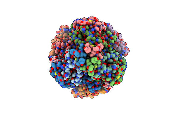
Organism: Homo sapiens
Method: ELECTRON MICROSCOPY Release Date: 2024-03-27 Classification: STRUCTURAL PROTEIN Ligands: CA, NAG |
 |
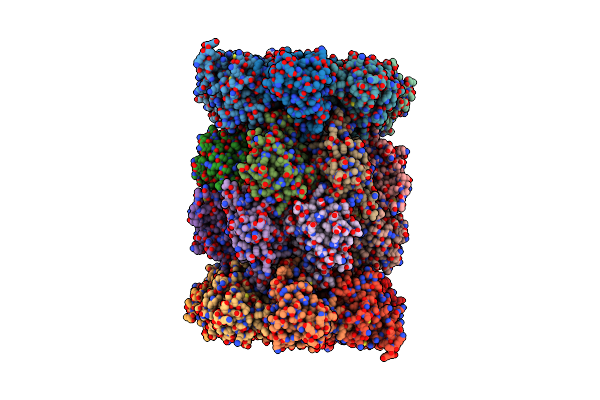
Organism: Homo sapiens
Method: X-RAY DIFFRACTION Resolution:1.50 Å Release Date: 2024-02-28 Classification: STRUCTURAL PROTEIN Ligands: ACT, NA, CA |
 |
Organism: Bacillus subtilis
Method: ELECTRON MICROSCOPY Release Date: 2022-01-05 Classification: CYTOSOLIC PROTEIN Ligands: FMN, F3S, FAD, SF4 |
 |
Glutamate Synthase, Glutamate Dehydrogenase Counter-Enzyme Complex (Gudb6-Glta6-Gltb6)
Organism: Bacillus subtilis subsp. subtilis ncib 3610 = atcc 6051 = dsm 10
Method: ELECTRON MICROSCOPY Release Date: 2022-01-05 Classification: CYTOSOLIC PROTEIN Ligands: FMN, F3S, FAD, SF4 |
 |
Structure Of The Membrane Soluble Spike Complex From The Lassa Virus In A C3-Symmetric Map
Organism: Lassa virus (strain mouse/sierra leone/josiah/1976)
Method: ELECTRON MICROSCOPY Release Date: 2021-12-29 Classification: VIRAL PROTEIN Ligands: NAG |
 |
Structure Of The Membrane Soluble Spike Complex From The Lassa Virus In A C1-Symmetric Map Focused On The Ectodomain
Organism: Lassa virus (strain mouse/sierra leone/josiah/1976)
Method: ELECTRON MICROSCOPY Release Date: 2021-12-29 Classification: VIRAL PROTEIN Ligands: NAG |
 |
Organism: Bacillus subtilis
Method: ELECTRON MICROSCOPY Release Date: 2021-04-07 Classification: DNA BINDING PROTEIN |
 |
Organism: Bacillus subtilis
Method: ELECTRON MICROSCOPY Release Date: 2021-04-07 Classification: DNA BINDING PROTEIN |
 |
Organism: Homo sapiens, Severe acute respiratory syndrome coronavirus 2
Method: ELECTRON MICROSCOPY Release Date: 2021-02-17 Classification: VIRUS Ligands: ZN, NAG |
 |
Organism: Homo sapiens
Method: ELECTRON MICROSCOPY Release Date: 2020-10-21 Classification: STRUCTURAL PROTEIN Ligands: CA, NAG |
 |
Organism: Homo sapiens
Method: ELECTRON MICROSCOPY Release Date: 2020-10-21 Classification: STRUCTURAL PROTEIN Ligands: CA, NAG |
 |
Organism: Rattus norvegicus
Method: ELECTRON MICROSCOPY Release Date: 2020-05-13 Classification: HYDROLASE |
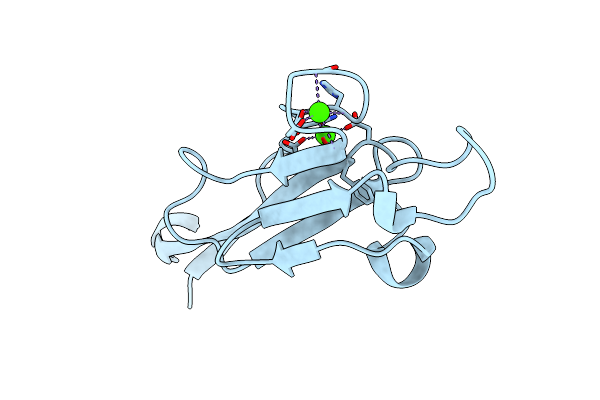 |
Organism: Homo sapiens
Method: X-RAY DIFFRACTION Resolution:1.63 Å Release Date: 2020-02-19 Classification: STRUCTURAL PROTEIN Ligands: CA |
 |
Organism: Homo sapiens, Ebola virus
Method: ELECTRON MICROSCOPY Release Date: 2020-02-12 Classification: VIRAL PROTEIN |
 |
Organism: Homo sapiens, Ebola virus
Method: ELECTRON MICROSCOPY Release Date: 2020-02-12 Classification: VIRAL PROTEIN |