
Search Count: 114
 |
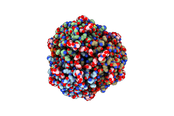
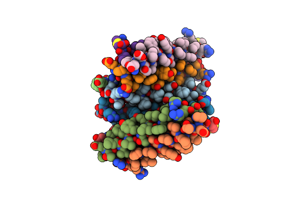
Organism: Mus musculus
Method: ELECTRON MICROSCOPY Release Date: 2024-02-14 Classification: VIRUS LIKE PARTICLE |
 |
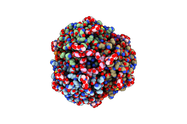
Organism: Mus musculus
Method: ELECTRON MICROSCOPY Release Date: 2024-02-14 Classification: VIRUS LIKE PARTICLE |
 |
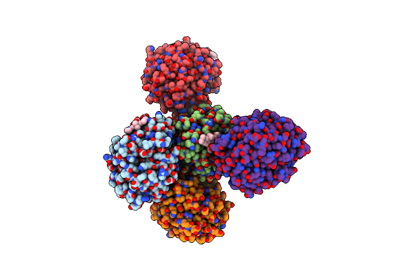
Organism: Mus musculus
Method: ELECTRON MICROSCOPY Release Date: 2024-02-14 Classification: VIRUS LIKE PARTICLE |
 |
Organism: Mus musculus
Method: ELECTRON MICROSCOPY Release Date: 2024-02-14 Classification: VIRUS LIKE PARTICLE |
 |
Structure Of The Membrane Soluble Spike Complex From The Lassa Virus In A C3-Symmetric Map
Organism: Lassa virus (strain mouse/sierra leone/josiah/1976)
Method: ELECTRON MICROSCOPY Release Date: 2021-12-29 Classification: VIRAL PROTEIN Ligands: NAG |
 |
Structure Of The Membrane Soluble Spike Complex From The Lassa Virus In A C1-Symmetric Map Focused On The Ectodomain
Organism: Lassa virus (strain mouse/sierra leone/josiah/1976)
Method: ELECTRON MICROSCOPY Release Date: 2021-12-29 Classification: VIRAL PROTEIN Ligands: NAG |
 |
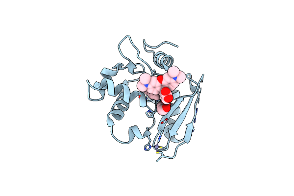
X-Ray Structure Of The Haloalkane Dehalogenase Hob (Halotag7-Based Oligonucleotide Binder) Labeled With A Chloroalkane-Tetramethylrhodamine Fluorophore Substrate
Organism: Rhodococcus sp.
Method: X-RAY DIFFRACTION Resolution:1.52 Å Release Date: 2021-04-21 Classification: HYDROLASE Ligands: OEH, ACT, CA |
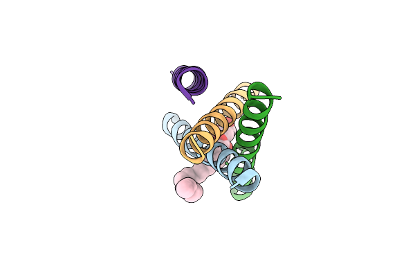 |
De Novo Designed Receptor Transmembrane Domains Enhance Car-T Cytotoxicity And Attenuate Cytokine Release
Organism: Synthetic construct
Method: X-RAY DIFFRACTION Resolution:2.55 Å Release Date: 2021-03-31 Classification: BIOSYNTHETIC PROTEIN Ligands: OLB |
 |
De Novo Designed Receptor Transmembrane Domains Enhance Car-T Cytotoxicity And Attenuate Cytokine Release
Organism: Synthetic construct
Method: X-RAY DIFFRACTION Resolution:2.70 Å Release Date: 2021-03-31 Classification: BIOSYNTHETIC PROTEIN Ligands: OLB |
 |
De Novo Designed Receptor Transmembrane Domains Enhance Car-T Cytotoxicity And Attenuate Cytokine Release
Organism: Synthetic construct
Method: X-RAY DIFFRACTION Resolution:3.48 Å Release Date: 2021-03-31 Classification: BIOSYNTHETIC PROTEIN |
 |
X-Ray Structure Of The Haloalkane Dehalogenase Halotag7 Labeled With A Chloroalkane-Tetramethylrhodamine Fluorophore Substrate
Organism: Rhodococcus sp.
Method: X-RAY DIFFRACTION Resolution:1.40 Å Release Date: 2021-03-31 Classification: HYDROLASE Ligands: OEH, CL, GOL |
 |
X-Ray Structure Of The Haloalkane Dehalogenase Halotag7 Labeled With A Chloroalkane-Carbopyronine Fluorophore Substrate
Organism: Rhodococcus sp.
Method: X-RAY DIFFRACTION Resolution:3.10 Å Release Date: 2021-03-31 Classification: HYDROLASE Ligands: OEK, CL |
 |
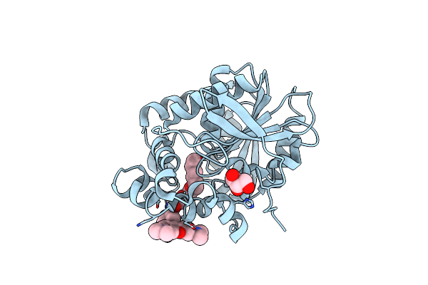
Crystal Structure Of Snap-Tag Labeled With A Benzyl-Tetramethylrhodamine Fluorophore
Organism: Homo sapiens
Method: X-RAY DIFFRACTION Resolution:2.30 Å Release Date: 2021-03-31 Classification: TRANSFERASE Ligands: ZN, OGQ, EDO |
 |
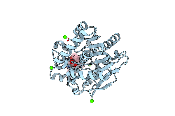
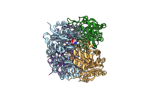
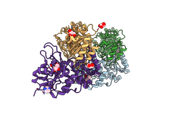
Organism: Escherichia coli (strain k12)
Method: X-RAY DIFFRACTION Resolution:1.75 Å Release Date: 2020-05-20 Classification: HYDROLASE |
 |
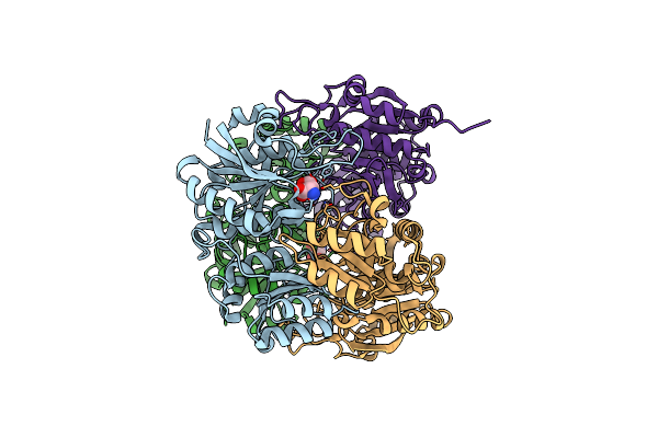
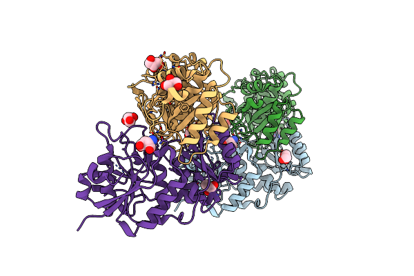
Complex Of Mutant (K162M) Of E. Coli L-Asparaginase Ii With L-Asp. Covalent Acyl-Enzyme Intermediate.
Organism: Escherichia coli (strain k12)
Method: X-RAY DIFFRACTION Resolution:1.90 Å Release Date: 2020-05-20 Classification: HYDROLASE Ligands: ASP |
 |
Organism: Escherichia coli (strain k12)
Method: X-RAY DIFFRACTION Resolution:1.78 Å Release Date: 2020-05-20 Classification: HYDROLASE Ligands: ASP |
 |
Organism: Escherichia coli (strain k12)
Method: X-RAY DIFFRACTION Resolution:1.80 Å Release Date: 2020-05-20 Classification: HYDROLASE Ligands: ASP |
 |
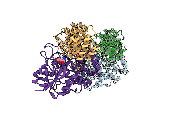
Complex Of Double Mutant (T89V,K162T) Of E. Coli L-Asparaginase Ii With L-Asp
Organism: Escherichia coli (strain k12)
Method: X-RAY DIFFRACTION Resolution:2.30 Å Release Date: 2020-05-20 Classification: HYDROLASE |
 |
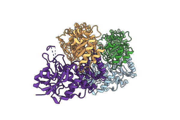
Complex Of Double Mutant (T89V,K162T) Of E. Coli L-Asparaginase Ii With L-Asp
Organism: Escherichia coli (strain k12)
Method: X-RAY DIFFRACTION Resolution:1.95 Å Release Date: 2020-05-20 Classification: HYDROLASE Ligands: IMD, GOL |
 |
Complex Of Double Mutant (T89V,K162T) Of E. Coli L-Asparaginase Ii With L-Asp
Organism: Escherichia coli (strain k12)
Method: X-RAY DIFFRACTION Resolution:2.00 Å Release Date: 2020-05-20 Classification: HYDROLASE Ligands: ASP, GOL |