
Search Count: 7
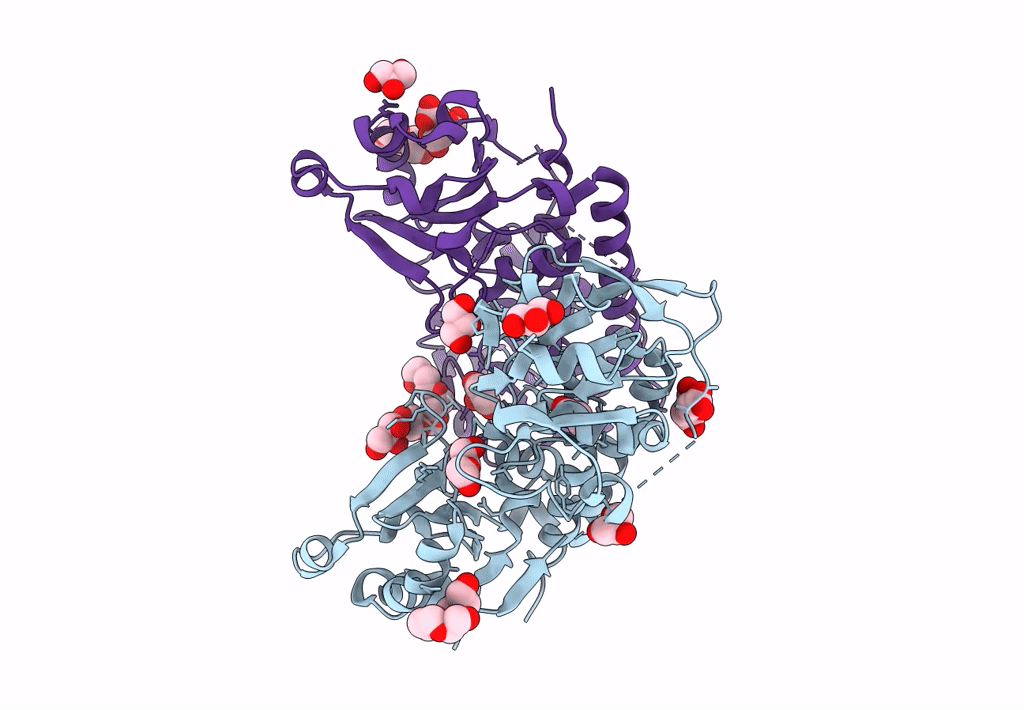 |
Putative Periplasmic Iron Siderophore Binding Protein Fecb (Rv3044) From Mycobacterium Tuberculosis
Organism: Mycobacterium tuberculosis h37rv
Method: X-RAY DIFFRACTION Resolution:2.00 Å Release Date: 2022-10-05 Classification: siderophore binding protein Ligands: GOL, CIT, PG4, 1PE |
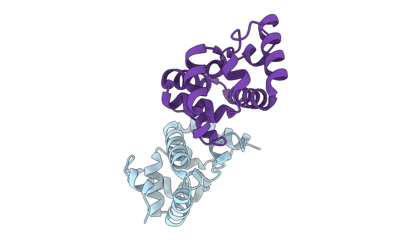 |
Organism: Mycolicibacterium smegmatis
Method: X-RAY DIFFRACTION Resolution:2.10 Å Release Date: 2021-08-18 Classification: UNKNOWN FUNCTION Ligands: BR |
 |
Structure Of Mycobacterium Tuberculosis Fructose 1,6-Bisphosphate Aldolase Bound To N-(4-Hydroxybutyl)- Glycolohydroxamic Acid Bis- Phosphate
Organism: Mycobacterium tuberculosis
Method: X-RAY DIFFRACTION Resolution:1.90 Å Release Date: 2011-10-12 Classification: LYASE Ligands: TD4, NA, ZN, SO4 |
 |
Structure Of Mycobacterium Tuberculosis Fructose 1,6-Bisphosphate Aldolase Bound To Sulfate
Organism: Mycobacterium tuberculosis
Method: X-RAY DIFFRACTION Resolution:2.35 Å Release Date: 2011-10-05 Classification: LYASE Ligands: SO4, NA |
 |
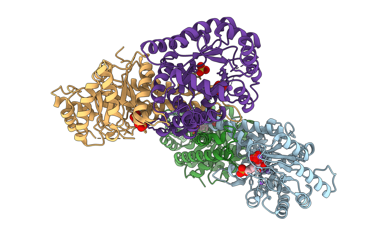
Crystal Structure Of Rv3671C From M. Tuberculosis H37Rv, Ser343Ala Mutant, Inactive Form
Organism: Mycobacterium tuberculosis h37rv
Method: X-RAY DIFFRACTION Resolution:2.10 Å Release Date: 2010-11-03 Classification: HYDROLASE |
 |
Organism: Mycobacterium tuberculosis
Method: X-RAY DIFFRACTION Resolution:1.30 Å Release Date: 2010-10-13 Classification: HYDROLASE |
 |
Organism: Mycobacterium tuberculosis
Method: X-RAY DIFFRACTION Resolution:1.75 Å Release Date: 2010-10-13 Classification: HYDROLASE |