
Search Count: 17
 |
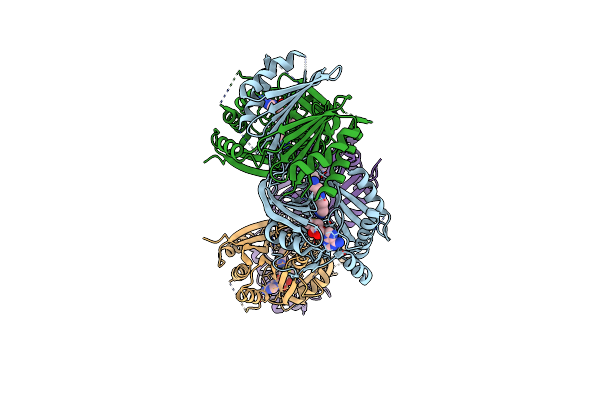
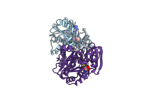
Organism: Escherichia coli (strain k12)
Method: X-RAY DIFFRACTION Resolution:1.85 Å Release Date: 2019-01-30 Classification: SIGNALING PROTEIN |
 |
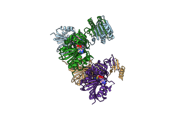
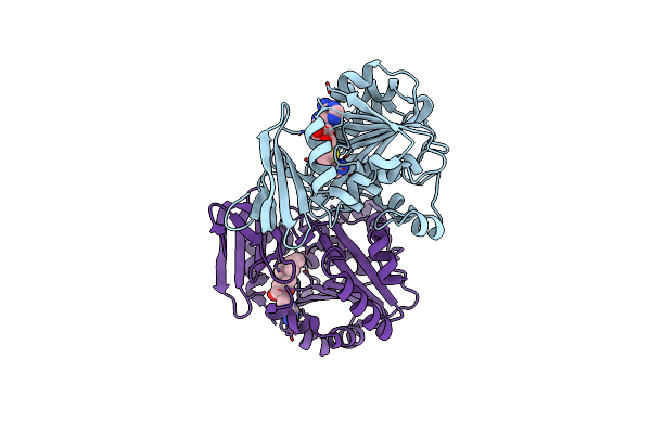
Organism: Escherichia coli (strain k12)
Method: X-RAY DIFFRACTION Resolution:2.70 Å Release Date: 2019-01-30 Classification: SIGNALING PROTEIN |
 |
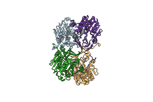
Organism: Escherichia coli
Method: X-RAY DIFFRACTION Resolution:1.33 Å Release Date: 2016-12-14 Classification: SIGNALING PROTEIN |
 |
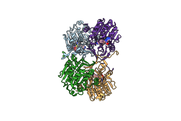
Organism: Escherichia coli
Method: X-RAY DIFFRACTION Resolution:2.30 Å Release Date: 2016-12-14 Classification: SIGNALING PROTEIN |
 |
Organism: Natronomonas pharaonis
Method: X-RAY DIFFRACTION Resolution:1.80 Å Release Date: 2011-08-31 Classification: MEMBRANE PROTEIN Ligands: RET, BNG, 22B |
 |
Organism: Natronomonas pharaonis
Method: X-RAY DIFFRACTION Resolution:2.10 Å Release Date: 2011-08-31 Classification: MEMBRANE PROTEIN Ligands: RET, BNG, 22B |
 |
Organism: Natronomonas pharaonis
Method: X-RAY DIFFRACTION Resolution:2.20 Å Release Date: 2011-08-31 Classification: MEMBRANE PROTEIN Ligands: RET, 22B, BR |
 |
Organism: Natronomonas pharaonis
Method: X-RAY DIFFRACTION Resolution:2.20 Å Release Date: 2011-08-31 Classification: MEMBRANE PROTEIN Ligands: RET, NO3, 22B |
 |
Organism: Natronomonas pharaonis dsm 2160
Method: X-RAY DIFFRACTION Resolution:2.00 Å Release Date: 2009-12-15 Classification: MEMBRANE PROTEIN Ligands: RET, 22B, L1P, L2P, L3P, CL |
 |
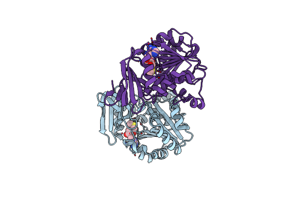
Crystal Structure Of Human Spermine Synthase In Complex With Spermidine And 5-Methylthioadenosine
Organism: Homo sapiens
Method: X-RAY DIFFRACTION Resolution:1.95 Å Release Date: 2008-02-19 Classification: TRANSFERASE Ligands: SPD, MTA |
 |
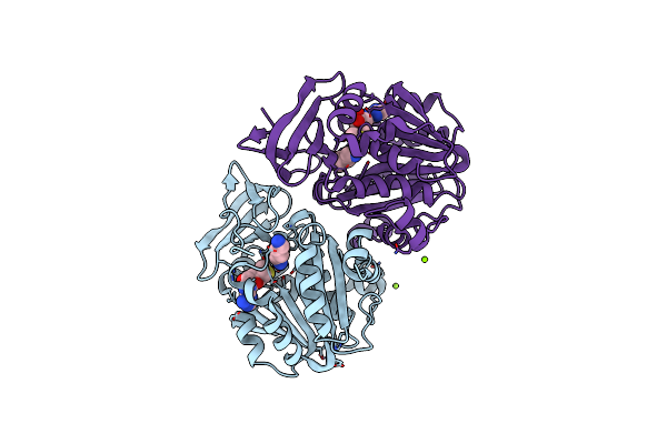
Crystal Structure Of Human Spermine Synthase In Complex With Spermine And 5-Methylthioadenosine
Organism: Homo sapiens
Method: X-RAY DIFFRACTION Resolution:2.45 Å Release Date: 2008-02-19 Classification: TRANSFERASE Ligands: SPM, MTA |
 |
Organism: Homo sapiens
Method: X-RAY DIFFRACTION Resolution:2.00 Å Release Date: 2006-12-12 Classification: TRANSFERASE Ligands: MTA |
 |
Organism: Homo sapiens
Method: X-RAY DIFFRACTION Resolution:2.00 Å Release Date: 2006-12-12 Classification: TRANSFERASE Ligands: MG, MTA, PUT |
 |
Organism: Homo sapiens
Method: X-RAY DIFFRACTION Resolution:1.89 Å Release Date: 2006-12-12 Classification: TRANSFERASE Ligands: SPD, MTA |
 |
Organism: Homo sapiens
Method: X-RAY DIFFRACTION Resolution:1.99 Å Release Date: 2006-12-12 Classification: TRANSFERASE Ligands: S4M |
 |
Organism: Thermotoga maritima
Method: X-RAY DIFFRACTION Resolution:1.50 Å Release Date: 2001-11-21 Classification: TRANSFERASE |
 |
Crystal Structure Of Spermidine Synthase In Complex With Transition State Analogue Adodato
Organism: Thermotoga maritima
Method: X-RAY DIFFRACTION Resolution:1.80 Å Release Date: 2001-11-21 Classification: TRANSFERASE Ligands: AAT |