
Search Count: 38
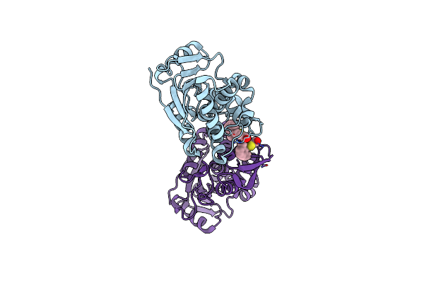 |
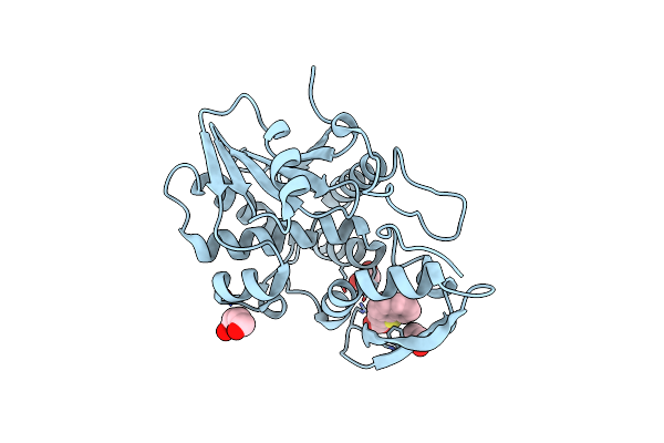
Organism: Ricinus communis
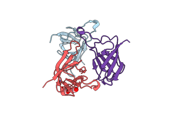
Method: X-RAY DIFFRACTION Resolution:1.80 Å Release Date: 2025-05-14 Classification: TOXIN,HYDROLASE/INHIBITOR Ligands: A1BH4, CL |
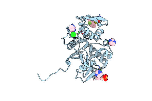 |
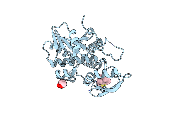
Organism: Ricinus communis
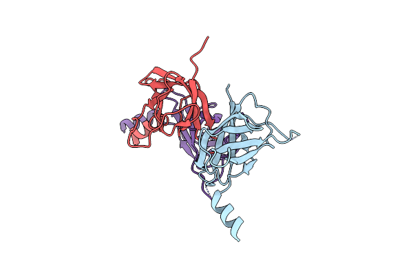
Method: X-RAY DIFFRACTION Resolution:1.80 Å Release Date: 2025-05-14 Classification: TOXIN,HYDROLASE/INHIBITOR Ligands: A1BH3, IMD, EPE, NI, CL |
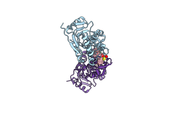 |
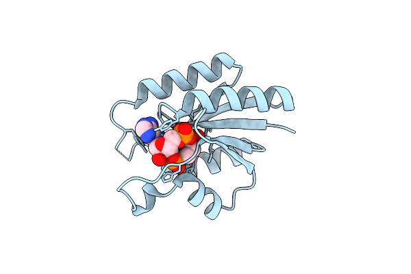
Organism: Ricinus communis
Method: X-RAY DIFFRACTION Resolution:1.80 Å Release Date: 2025-05-14 Classification: TOXIN,HYDROLASE/INHIBITOR Ligands: A1BH2, EDO |
 |
Solution Structure Of A Parallel Stranded G-Quadruplex Formed In Orai1 Promoter
|
 |
Organism: Ricinus communis
Method: X-RAY DIFFRACTION Resolution:1.95 Å Release Date: 2024-05-01 Classification: TOXIN Ligands: ZJT, 2PE |
 |
Organism: Ricinus communis
Method: X-RAY DIFFRACTION Resolution:2.26 Å Release Date: 2024-05-01 Classification: TOXIN,HYDROLASE/INHIBITOR Ligands: CL, U4T, EDO, 2PE |
 |
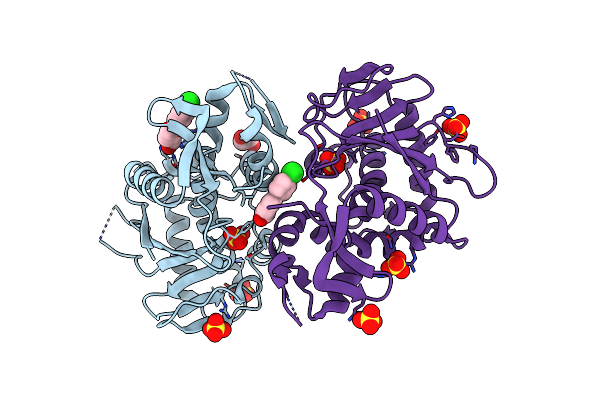
Organism: Ricinus communis
Method: X-RAY DIFFRACTION Resolution:2.76 Å Release Date: 2024-05-01 Classification: TOXIN,HYDROLASE/INHIBITOR Ligands: ZXJ, CL, 2PE |
 |
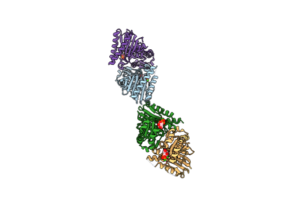
Organism: Escherichia coli
Method: X-RAY DIFFRACTION Resolution:1.91 Å Release Date: 2024-03-06 Classification: TOXIN Ligands: P1U, EDO, CL, SO4 |
 |
Organism: Escherichia coli
Method: X-RAY DIFFRACTION Resolution:3.10 Å Release Date: 2024-03-06 Classification: TOXIN |
 |
Crystal Structure Of The Mlad Domain Of The Mlad Protein From Escherichia Coli (Form I)
Organism: Escherichia coli k-12
Method: X-RAY DIFFRACTION Resolution:2.30 Å Release Date: 2024-01-10 Classification: TRANSPORT PROTEIN Ligands: CO2, EDO |
 |
Crystal Structure Of The Mlad Domain Of The Mlad Protein From Escherichia Coli (Form Ii)
Organism: Escherichia coli k-12
Method: X-RAY DIFFRACTION Resolution:2.70 Å Release Date: 2024-01-10 Classification: TRANSPORT PROTEIN Ligands: MG, CO2 |
 |
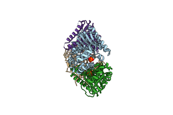
Crystal Structure Of The Ectodomain Of The Mlad Protein From Escherichia Coli In The Resting State
Organism: Escherichia coli k-12
Method: X-RAY DIFFRACTION Resolution:3.20 Å Release Date: 2024-01-10 Classification: TRANSPORT PROTEIN |
 |
Organism: Escherichia coli k-12
Method: X-RAY DIFFRACTION Resolution:2.50 Å Release Date: 2022-09-21 Classification: TRANSPORT PROTEIN Ligands: PEF, EDO |
 |
Crystal Structure Of Crispr-Associated Transcription Factor Csa3 Complexed With Ca4
Organism: Saccharolobus solfataricus, Synthetic construct
Method: X-RAY DIFFRACTION Resolution:2.05 Å Release Date: 2021-11-17 Classification: TRANSCRIPTION Ligands: GOL |
 |
Universal Stress Protein (Usp) Domain Of Kdpd Histidine Kinase In Complex With Second Messenger C-Di-Amp
Organism: Staphylococcus aureus subsp. aureus mrsa252
Method: X-RAY DIFFRACTION Resolution:2.30 Å Release Date: 2021-05-26 Classification: SIGNALING PROTEIN Ligands: 2BA |
 |
Organism: Rattus norvegicus
Method: X-RAY DIFFRACTION Resolution:2.51 Å Release Date: 2018-12-19 Classification: MEMBRANE PROTEIN Ligands: NAG |
 |
Organism: Rattus norvegicus
Method: X-RAY DIFFRACTION Resolution:1.96 Å Release Date: 2018-12-19 Classification: MEMBRANE PROTEIN Ligands: NAG, PO4, DMS, GOL |
 |
Organism: Staphylococcus aureus (strain mssa476)
Method: X-RAY DIFFRACTION Resolution:2.40 Å Release Date: 2017-02-08 Classification: HYDROLASE Ligands: CA, IPD, PO4 |
 |
Organism: Staphylococcus aureus (strain mssa476)
Method: X-RAY DIFFRACTION Resolution:2.20 Å Release Date: 2016-10-05 Classification: HYDROLASE Ligands: PO4 |
 |
Organism: Staphylococcus aureus
Method: X-RAY DIFFRACTION Resolution:2.20 Å Release Date: 2015-12-23 Classification: HYDROLASE Ligands: CA, PGE, GOL, PG0, NAP, PG4, CL |