
Search Count: 24
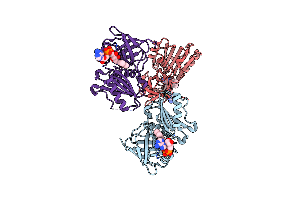 |
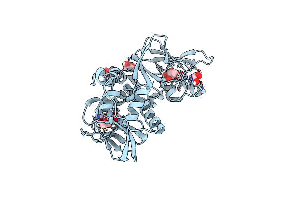
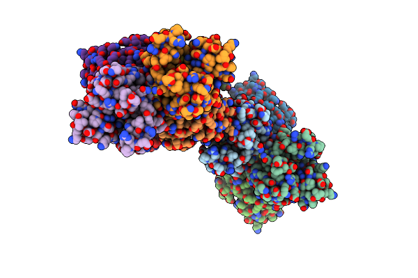
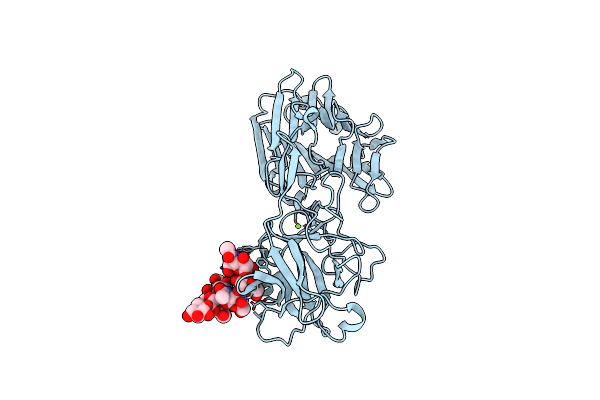
Spnox Dehydrogenase Domain, Mutant F397W In Complex With Flavin Adenine Dinucleotide (Fad)
Organism: Streptococcus pneumoniae
Method: X-RAY DIFFRACTION Resolution:1.94 Å Release Date: 2024-05-08 Classification: MEMBRANE PROTEIN Ligands: FAD, BR |
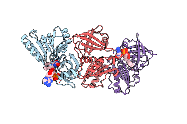 |
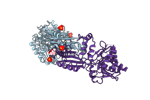
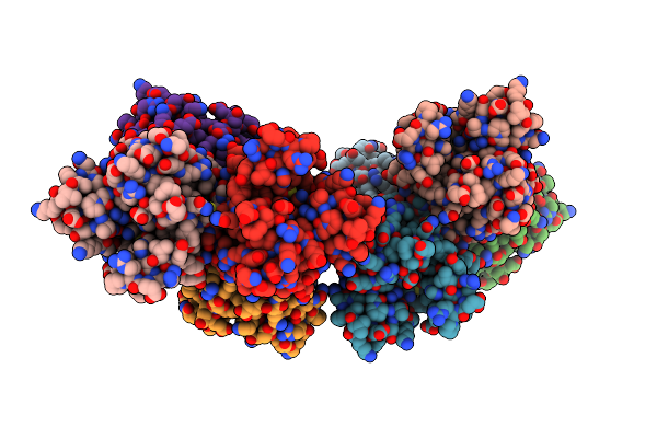
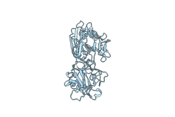
Organism: Streptococcus pneumoniae
Method: X-RAY DIFFRACTION Resolution:2.50 Å Release Date: 2024-05-08 Classification: ELECTRON TRANSPORT Ligands: FAD, CL |
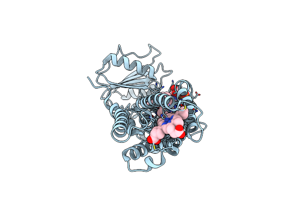 |
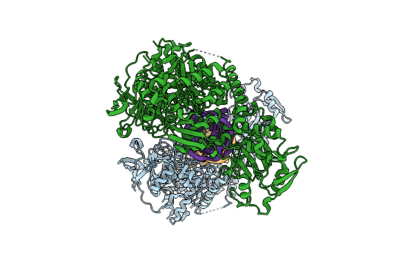
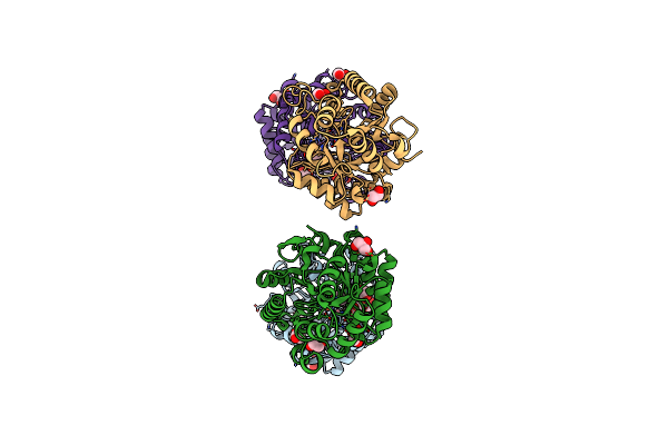
Organism: Streptococcus pneumoniae
Method: X-RAY DIFFRACTION Resolution:3.62 Å Release Date: 2024-05-08 Classification: ELECTRON TRANSPORT Ligands: FDA, HEM |
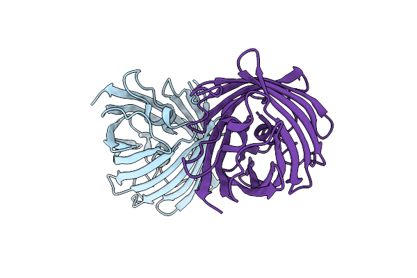 |
Organism: Discosoma sp.
Method: X-RAY DIFFRACTION Resolution:1.33 Å Release Date: 2023-04-05 Classification: FLUORESCENT PROTEIN |
 |
Organism: Branchiostoma lanceolatum
Method: X-RAY DIFFRACTION Resolution:1.51 Å Release Date: 2022-05-04 Classification: FLUORESCENT PROTEIN |
 |
Organism: Branchiostoma lanceolatum
Method: X-RAY DIFFRACTION Resolution:1.95 Å Release Date: 2022-05-04 Classification: FLUORESCENT PROTEIN Ligands: SO4 |
 |
Structure Of The T207D Single-Point Mutant Of The Fluorescent Protein Neoncyan1 At Ph 6.5
Organism: Branchiostoma lanceolatum
Method: X-RAY DIFFRACTION Resolution:1.60 Å Release Date: 2022-05-04 Classification: FLUORESCENT PROTEIN |
 |
Regulatory Subunit Of A Camp-Independent Protein Kinase A From Trypanosoma Cruzi At 1.09 A Resolution
Organism: Trypanosoma cruzi (strain cl brener)
Method: X-RAY DIFFRACTION Resolution:1.09 Å Release Date: 2019-04-03 Classification: SIGNALING PROTEIN Ligands: EDO, 7CI |
 |
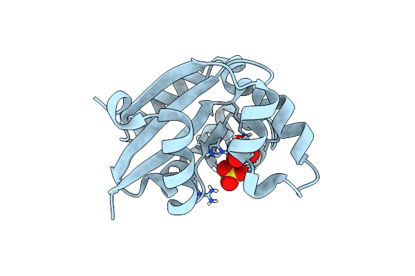
The Crystal Structure Of A M20 Family Metallo-Carboxypeptidase Sso-Cp2 From Sulfolobus Solfataricus
Organism: Sulfolobus solfataricus
Method: X-RAY DIFFRACTION Resolution:2.34 Å Release Date: 2014-10-15 Classification: HYDROLASE Ligands: ZN, SO4, GOL |
 |
Orthorhombic Crystal Form C222 Of The Aquifex Aeolicus Nucleoside Diphosphate Kinase
Organism: Aquifex aeolicus
Method: X-RAY DIFFRACTION Resolution:1.47 Å Release Date: 2012-03-14 Classification: TRANSFERASE Ligands: SO4 |
 |
Orthorhombic Crystal Form P21212 Of The Aquifex Aeolicus Nucleoside Diphosphate Kinase
Organism: Aquifex aeolicus
Method: X-RAY DIFFRACTION Resolution:1.37 Å Release Date: 2012-03-14 Classification: TRANSFERASE Ligands: GOL, SO4 |
 |
Hexagonal Form P6122 Of The Aquifex Aeolicus Nucleoside Diphosphate Kinase (First Stage Of Radiation Damage)
Organism: Aquifex aeolicus
Method: X-RAY DIFFRACTION Resolution:2.30 Å Release Date: 2012-03-14 Classification: TRANSFERASE |
 |
Hexagonal Form P6122 Of The Aquifex Aeolicus Nucleoside Diphosphate Kinase (Final Stage Of Radiation Damage)
Organism: Aquifex aeolicus
Method: X-RAY DIFFRACTION Resolution:2.30 Å Release Date: 2012-03-14 Classification: TRANSFERASE |
 |
Hexagonal Crystal Form P61 Of The Aquifex Aeolicus Nucleoside Diphosphate Kinase
Organism: Aquifex aeolicus
Method: X-RAY DIFFRACTION Resolution:2.10 Å Release Date: 2012-02-29 Classification: TRANSFERASE |
 |
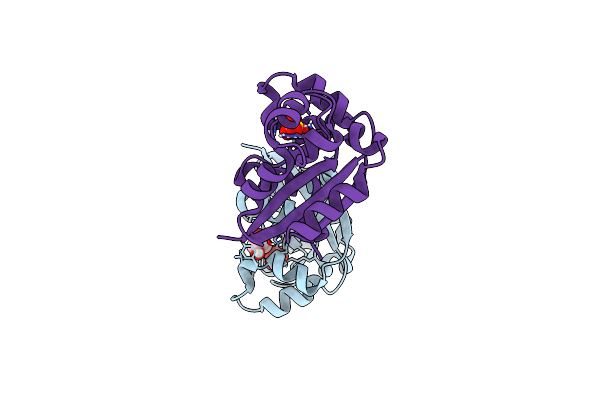
Crystal Structure Analysis Of Iron Regulatory Protein 1 In Complex With Ferritin H Ire Rna
Organism: Oryctolagus cuniculus
Method: X-RAY DIFFRACTION Resolution:2.80 Å Release Date: 2011-07-27 Classification: LYASE/RNA |
 |
Organism: Clostridium botulinum
Method: X-RAY DIFFRACTION Resolution:1.90 Å Release Date: 2009-07-07 Classification: TOXIN Ligands: SO4, GOL |
 |
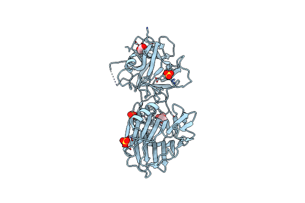
Crystal Structure Of Botulinum Neurotoxin Serotype A Binding Domain In Complex With Gt1B
Organism: Clostridium botulinum
Method: X-RAY DIFFRACTION Resolution:1.60 Å Release Date: 2008-08-26 Classification: HYDROLASE Ligands: MG |
 |
Organism: Clostridium botulinum
Method: X-RAY DIFFRACTION Resolution:1.70 Å Release Date: 2008-08-26 Classification: HYDROLASE |
 |
Structural Basis For Natural Lactonase And Promiscuous Phosphotriesterase Activities
Organism: Sulfolobus solfataricus
Method: X-RAY DIFFRACTION Resolution:2.60 Å Release Date: 2008-04-15 Classification: HYDROLASE Ligands: FE2, CO, EDO, GOL |
 |
Structural Basis For Natural Lactonase And Promiscuous Phosphotriesterase Activities
Organism: Sulfolobus solfataricus
Method: X-RAY DIFFRACTION Resolution:2.05 Å Release Date: 2008-04-15 Classification: HYDROLASE Ligands: FE2, CO, GOL, EDO, HT5 |