
Search Count: 5
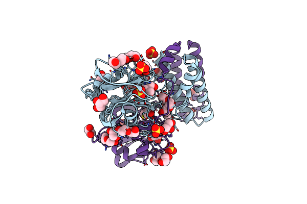 |
Organism: Streptococcus pneumoniae
Method: X-RAY DIFFRACTION Resolution:1.39 Å Release Date: 2013-08-07 Classification: HYDROLASE Ligands: EDO, TRS, SO4, PEG, GOL, PGE |
 |
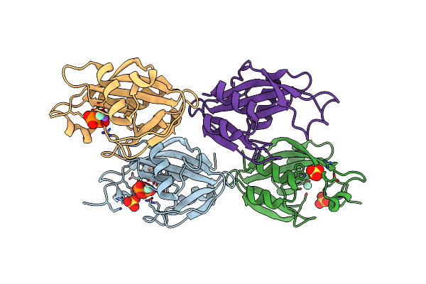
Organism: Bacillus cereus
Method: X-RAY DIFFRACTION Resolution:1.80 Å Release Date: 2011-07-13 Classification: HYDROLASE Ligands: SO4 |
 |
Organism: Bacillus cereus
Method: X-RAY DIFFRACTION Resolution:2.50 Å Release Date: 2011-07-13 Classification: HYDROLASE Ligands: GD3 |
 |
Structure Of The E. Coli Dihydroneopterin Triphosphate Pyrophosphohydrolase
Organism: Escherichia coli
Method: X-RAY DIFFRACTION Resolution:1.80 Å Release Date: 2007-08-28 Classification: HYDROLASE Ligands: SO4, PPV |
 |
Structure Of The E. Coli Dihydroneopterin Triphosphate Pyrophosphohydrolase In Complex With Sm+3 And Pyrophosphate
Organism: Escherichia coli
Method: X-RAY DIFFRACTION Resolution:2.60 Å Release Date: 2007-08-28 Classification: HYDROLASE Ligands: SO4, SM, NA, PPV |