
Search Count: 4
 |
Crystal Structure Of Biotin Carboxylase From E.Coli In Complex With Potent Inhibitor 1
Organism: Escherichia coli
Method: X-RAY DIFFRACTION Resolution:2.10 Å Release Date: 2009-01-13 Classification: LIGASE Ligands: LZJ, CL |
 |
Crystal Structure Of Biotin Carboxylase From E.Coli In Complex With Potent Inhibitor 2
Organism: Escherichia coli
Method: X-RAY DIFFRACTION Resolution:2.40 Å Release Date: 2009-01-13 Classification: LIGASE Ligands: LZK |
 |
Crystal Structure Of Biotin Carboxylase From E.Coli In Complex With Potent Inhibitor 3
Organism: Escherichia coli
Method: X-RAY DIFFRACTION Resolution:2.31 Å Release Date: 2009-01-13 Classification: LIGASE Ligands: LZL, CL |
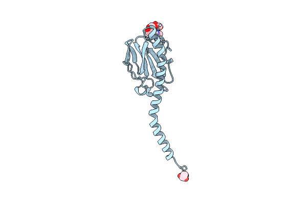 |
Crystallographic Structure Of Phosphorylated Pilin From Neisseria: Phosphoserine Sites Modify Type Iv Pilus Surface Chemistry
Organism: Neisseria gonorrhoeae
Method: X-RAY DIFFRACTION Resolution:2.60 Å Release Date: 1998-05-27 Classification: CELL ADHESION Ligands: PT, HTO |