
Search Count: 15
 |
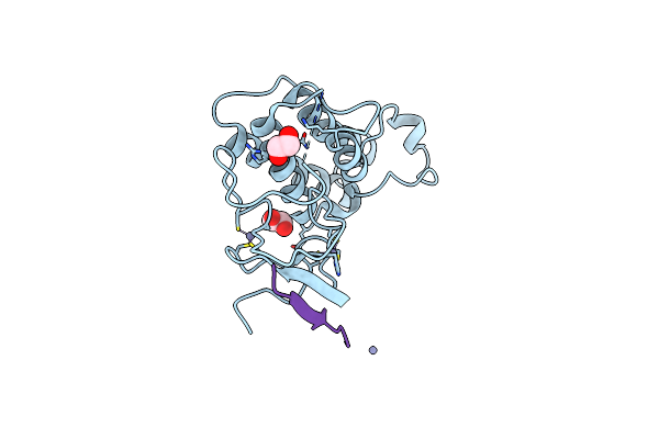
Crystal Structure Of Idh1 Variant R132C S280F In Complex With Nadph, Ca2+ And 3-Butyl-2-Oxoglutarate
Organism: Homo sapiens
Method: X-RAY DIFFRACTION Resolution:2.35 Å Release Date: 2022-11-02 Classification: OXIDOREDUCTASE Ligands: GOL, NDP, QDC, QD8, CA |
 |
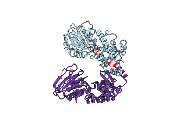
Crystal Structure Of Trim33 Phd-Bromodomain Isoform B In Complex With H3K10Ac Histone Peptide.
Organism: Homo sapiens
Method: X-RAY DIFFRACTION Resolution:1.63 Å Release Date: 2022-06-29 Classification: TRANSCRIPTION Ligands: EDO, ZN |
 |
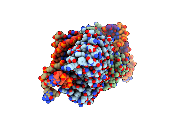
Structure Of Keap1 Kelch Domain With (1S,2R)-2-{[(1S)-1-[(1,3-Dioxo-1,3-Dihydro-2H-Isoindol-2-Yl)Methyl]-3,4-Dihydroisoquinolin-2(1H)-Yl]Carbonyl}Cyclohexanecarboxylic Acid
Organism: Homo sapiens
Method: X-RAY DIFFRACTION Resolution:2.41 Å Release Date: 2014-02-19 Classification: TRANSCRIPTION/INHIBITOR Ligands: ACT, 1VV, NA |
 |
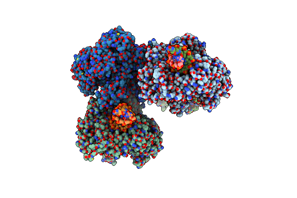
Structure Of Keap1 Kelch Domain With 2-{[(1S)-2-{[(1R,2S)-2-(1H-Tetrazol-5-Yl)Cyclohexyl]Carbonyl}-1,2,3,4-Tetrahydroisoquinolin-1-Yl]Methyl}-1H-Isoindole-1,3(2H)-Dione
Organism: Homo sapiens
Method: X-RAY DIFFRACTION Resolution:2.40 Å Release Date: 2014-02-19 Classification: TRANSCRIPTION/INHIBITOR Ligands: 1VW, ACT |
 |
Structure Of Keap1 Kelch Domain With (1S,2R)-2-{[(1S)-5-Methyl-1-[(1-Oxo-1,3-Dihydro-2H-Isoindol-2-Yl)Methyl]-3,4-Dihydroisoquinolin-2(1H)-Yl]Carbonyl}Cyclohexanecarboxylic Acid
Organism: Homo sapiens
Method: X-RAY DIFFRACTION Resolution:2.25 Å Release Date: 2014-02-19 Classification: TRANSCRIPTION/INHIBITOR Ligands: 1VX, ACT |
 |
Structure Of Keap1 Kelch Domain With(1S,2R)-2-[(1S)-1-[(1-Oxo-2,3-Dihydro-1H-Isoindol-2-Yl)Methyl]-1,2,3,4-Tetrahydroisoquinoline-2-Carbonyl]Cyclohexane-1-Carboxylic Acid
Organism: Homo sapiens
Method: X-RAY DIFFRACTION Resolution:2.55 Å Release Date: 2014-02-19 Classification: TRANSCRIPTION/INHIBITOR Ligands: 2FS, ACT |
 |
Organism: Arabidopsis thaliana
Method: X-RAY DIFFRACTION Resolution:2.30 Å Release Date: 2012-07-25 Classification: HYDROLASE Ligands: GOL |
 |
Organism: Arabidopsis thaliana
Method: X-RAY DIFFRACTION Resolution:1.70 Å Release Date: 2012-07-25 Classification: HYDROLASE Ligands: GOL |
 |
Organism: Arabidopsis thaliana
Method: X-RAY DIFFRACTION Resolution:2.80 Å Release Date: 2012-07-25 Classification: HYDROLASE/DNA |
 |
Crystal Structure Of A Replicative Dna Polymerase Bound To Dna Containing Thymine Glycol
Organism: Enterobacteria phage rb69
Method: X-RAY DIFFRACTION Resolution:2.84 Å Release Date: 2011-08-10 Classification: TRANSFERASE/DNA |
 |
Crystal Structure Of A Replicative Dna Polymerase Bound To Dna Containing Thymine Glycol
Organism: Enterobacteria phage rb69
Method: X-RAY DIFFRACTION Resolution:2.65 Å Release Date: 2011-08-10 Classification: TRANSFERASE/DNA Ligands: SO4 |
 |
Crystal Structure Of A Replicative Dna Polymerase Bound To Dna Containing Thymine Glycol
Organism: Enterobacteria phage rb69
Method: X-RAY DIFFRACTION Resolution:3.00 Å Release Date: 2011-08-10 Classification: TRANSFERASE/DNA |
 |
Crystal Structure Of A Replicative Dna Polymerase Bound To Dna Containing Thymine Glycol
Organism: Enterobacteria phage rb69
Method: X-RAY DIFFRACTION Resolution:2.98 Å Release Date: 2011-08-10 Classification: TRANSFERASE/DNA Ligands: DTP |
 |
Crystal Structure Of Methanocaldococcus Jannaschii 8-Oxoguanine Dna Glycosylase (Mjogg)
Organism: Methanocaldococcus jannaschii
Method: X-RAY DIFFRACTION Resolution:2.00 Å Release Date: 2009-05-19 Classification: DNA repair, HYDROLASE, LYASE |
 |
Crystal Structure Of Sulfolobus Solfataricus 8-Oxoguanine Dna Glycosylase (Ssogg)
Organism: Sulfolobus solfataricus
Method: X-RAY DIFFRACTION Resolution:1.90 Å Release Date: 2009-05-19 Classification: DNA repair, HYDROLASE, LYASE Ligands: GOL, SO4 |