
Search Count: 240
 |
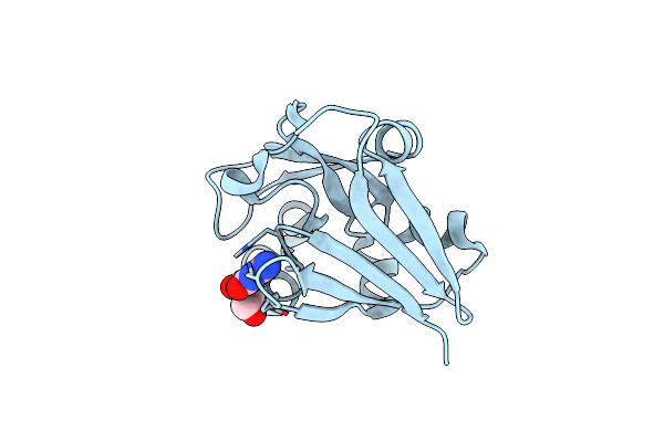
Crystal Structure Of Sh3-Like_Bac-Type Domain (79-145) Of Conserved Domain Protein Gbaa_2967 From Bacillus Anthracis Ames Ancestor
Organism: Bacillus anthracis str. ames
Method: X-RAY DIFFRACTION Release Date: 2025-06-11 Classification: UNKNOWN FUNCTION Ligands: MG, FMT |
 |
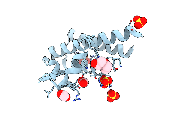
Crystal Structure Of Nlpc/P60 Domain From Clostridium Innocuum Nlpc/P60 Domain-Containing Protein Ci_01448.
Organism: [clostridium] innocuum 2959
Method: X-RAY DIFFRACTION Resolution:1.92 Å Release Date: 2023-10-18 Classification: HYDROLASE Ligands: SO4, EDO, GOL |
 |
Crystal Structure Of The Hypothetical Protein (Acx60_00475) From Acinetobacter Baumannii
Organism: Acinetobacter baumannii
Method: X-RAY DIFFRACTION Resolution:1.65 Å Release Date: 2022-11-30 Classification: UNKNOWN FUNCTION Ligands: PEG, EDO |
 |
Crystal Structure Of Putative Pterin Binding Protein (Prur) From Klebsiella Pneumoniae In Complex With Neopterin
Organism: Klebsiella pneumoniae subsp. pneumoniae sa1
Method: X-RAY DIFFRACTION Resolution:2.50 Å Release Date: 2022-08-10 Classification: PTERIN BINDING PROTEIN Ligands: NEU, CL, SO4 |
 |
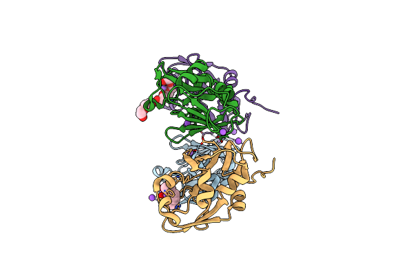
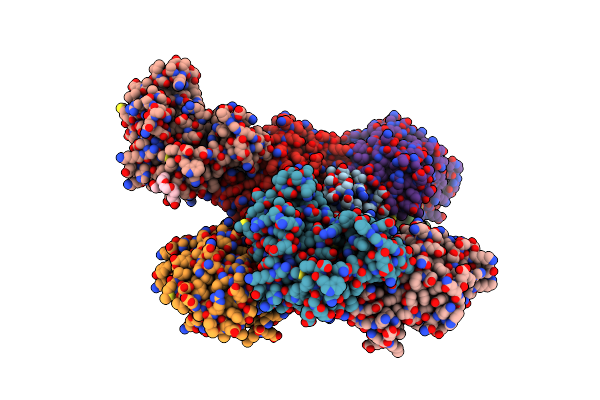
Crystal Structure Of Putataive Short-Chain Dehydrogenase/Reductase (Fabg) From Klebsiella Pneumoniae Subsp. Pneumoniae Ntuh-K2044 In Complex With Nadh
Organism: Klebsiella pneumoniae subsp. pneumoniae ntuh-k2044
Method: X-RAY DIFFRACTION Resolution:2.60 Å Release Date: 2022-03-02 Classification: BIOSYNTHETIC PROTEIN Ligands: K, CL, NAI, GOL, EDO |
 |
Organism: Klebsiella pneumoniae subsp. pneumoniae ntuh-k2044
Method: X-RAY DIFFRACTION Resolution:2.69 Å Release Date: 2022-02-02 Classification: HYDROLASE Ligands: EDO |
 |
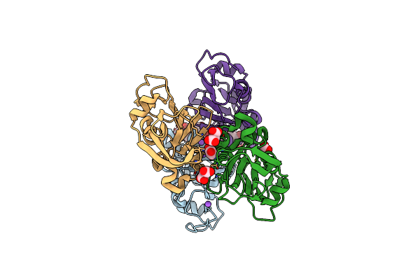
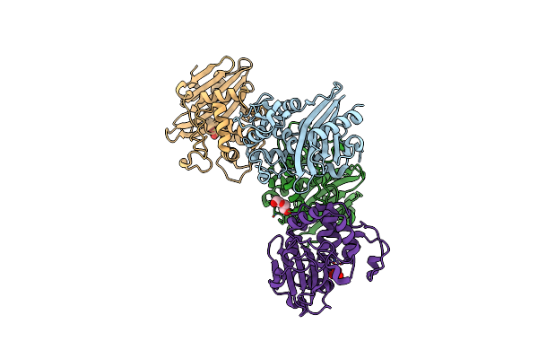
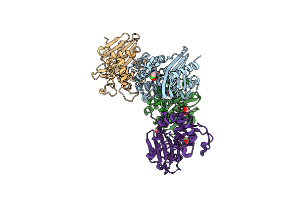
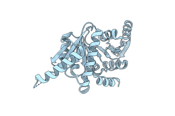
Crystal Structure Of The Succinyl-Diaminopimelate Desuccinylase (Dape) From Acinetobacter Baumannii In Complex With Succinic Acid
Organism: Acinetobacter baumannii atcc 17978
Method: X-RAY DIFFRACTION Resolution:2.25 Å Release Date: 2021-12-15 Classification: HYDROLASE Ligands: SIN, ACT, PGE, ZN |
 |
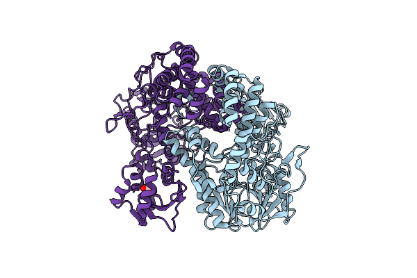
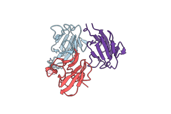
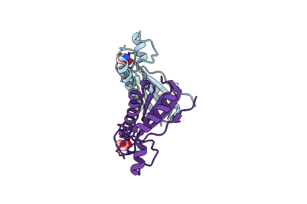
Crystal Structure Of The Dna-Binding Transcriptional Repressor Deor From Escherichia Coli Str. K-12
Organism: Escherichia coli (strain k12)
Method: X-RAY DIFFRACTION Resolution:1.75 Å Release Date: 2021-12-01 Classification: DNA BINDING PROTEIN Ligands: EDO, SO4, CL |
 |
Crystal Structure Of Peptidyl-Prolyl Cis-Trans Isomerasefrom (Ppib) Streptococcus Pneumoniae R6
Organism: Streptococcus pneumoniae (strain atcc baa-255 / r6)
Method: X-RAY DIFFRACTION Resolution:1.88 Å Release Date: 2021-12-01 Classification: ISOMERASE Ligands: CL, EDO, MES |
 |
High Resolution Crystal Structure Of Putative Pterin Binding Protein Prur (Vv2_1280) From Vibrio Vulnificus Cmcp6
Organism: Vibrio vulnificus (strain cmcp6)
Method: X-RAY DIFFRACTION Resolution:0.99 Å Release Date: 2021-11-17 Classification: Pterin binding protein Ligands: FMT, NA |
 |
1.50 Angstroms Resolution Crystal Structure Of Putative Pterin Binding Protein Prur (Atu3496) From Agrobacterium Fabrum Str. C58
Organism: Agrobacterium fabrum (strain c58 / atcc 33970)
Method: X-RAY DIFFRACTION Resolution:1.50 Å Release Date: 2021-11-17 Classification: Pterin binding protein |
 |
1.83 Angstroms Resolution Crystal Structure Of Putative Pterin Binding Protein Prur (Atu3496) From Agrobacterium Fabrum Str. C58
Organism: Agrobacterium fabrum (strain c58 / atcc 33970)
Method: X-RAY DIFFRACTION Resolution:1.83 Å Release Date: 2021-11-17 Classification: Pterin binding protein Ligands: CL, NA, PG4, PEG |
 |
High Resolution Crystal Structure Of Putative Pterin Binding Protein (Prur) From Vibrio Cholerae O1 Biovar El Tor Str. N16961 In Complex With Neopterin
Organism: Vibrio cholerae o1 biovar el tor str. n16961
Method: X-RAY DIFFRACTION Resolution:1.03 Å Release Date: 2021-11-17 Classification: PTERIN-BINDING PROTEIN Ligands: NEU |
 |
Crystal Structure Of C79A Mutant Of Class D Beta-Lactamase From Clostridium Difficile 630
Organism: Clostridioides difficile
Method: X-RAY DIFFRACTION Resolution:1.95 Å Release Date: 2021-08-11 Classification: HYDROLASE Ligands: PEG, SO4 |
 |
Crystal Structure Of K83A Mutant Of Class D Beta-Lactamase From Clostridium Difficile 630
Organism: Clostridioides difficile
Method: X-RAY DIFFRACTION Resolution:1.88 Å Release Date: 2021-08-11 Classification: HYDROLASE Ligands: CL, ACT, EDO, NA |
 |
Crystal Structure Of The Forkhead Associated (Fha) Domain Of The Glycogen Accumulation Regulator (Gara) From Mycobacterium Tuberculosis
Organism: Mycobacterium tuberculosis
Method: X-RAY DIFFRACTION Resolution:1.68 Å Release Date: 2021-07-28 Classification: SIGNALING PROTEIN Ligands: PEG |
 |
Crystal Structure Of The Peptidoglycan Binding Domain Of The Outer Membrane Protein (Ompa) From Klebsiella Pneumoniae With Bound D-Alanine
Organism: Klebsiella pneumoniae subsp. pneumoniae
Method: X-RAY DIFFRACTION Resolution:1.88 Å Release Date: 2021-07-28 Classification: PEPTIDE BINDING PROTEIN Ligands: DAL, CL |
 |
Crystal Structure Of Putative Universal Stress Protein From Pseudomonas Aeruginosa Ucbpp-Pa14
Organism: Pseudomonas aeruginosa ucbpp-pa14
Method: X-RAY DIFFRACTION Resolution:1.25 Å Release Date: 2021-05-19 Classification: UNKNOWN FUNCTION Ligands: CL |
 |
1.90 Angstrom Resolution Crystal Structure Phosphoadenosine Phosphosulfate Reductase (Cysh) From Vibrio Vulnificus
Organism: Vibrio vulnificus (strain cmcp6)
Method: X-RAY DIFFRACTION Resolution:1.90 Å Release Date: 2021-02-10 Classification: OXIDOREDUCTASE Ligands: PG4, PEG, P6G |
 |
High Resolution Crystal Structure Of The Dna-Binding Domain From The Sensor Histidine Kinase Chis From Vibrio Cholerae
Organism: Vibrio cholerae o1 biovar el tor
Method: X-RAY DIFFRACTION Resolution:1.28 Å Release Date: 2020-11-25 Classification: DNA BINDING PROTEIN Ligands: SO4, PEG, FMT |