
Search Count: 97
 |
Organism: Homo sapiens
Method: X-RAY DIFFRACTION Release Date: 2025-12-03 Classification: UNKNOWN FUNCTION |
 |
Organism: Homo sapiens
Method: X-RAY DIFFRACTION Release Date: 2025-12-03 Classification: UNKNOWN FUNCTION |
 |
Organism: Escherichia coli, Escherichia phage t7
Method: ELECTRON MICROSCOPY Release Date: 2024-12-11 Classification: TRANSFERASE |
 |
Organism: Arabidopsis thaliana
Method: ELECTRON MICROSCOPY Release Date: 2024-04-03 Classification: TRANSFERASE Ligands: FE |
 |
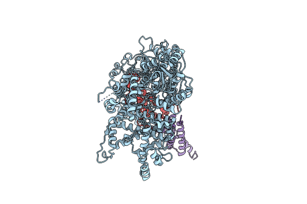
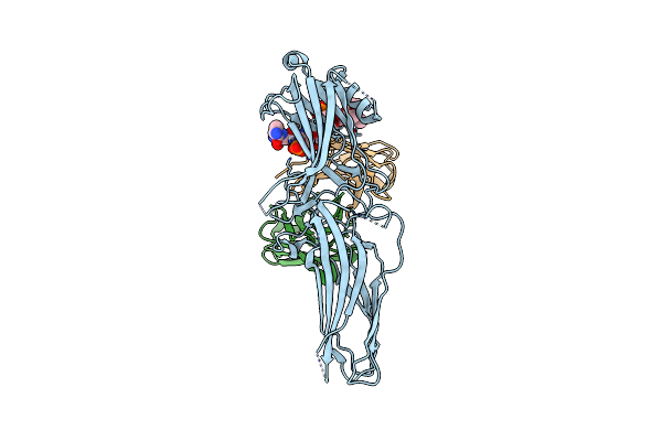
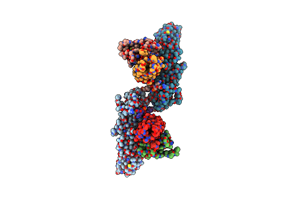
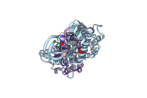
Structure Of Beta-Arrestin2 In Complex With A Phosphopeptide Corresponding To The Human Atypical Chemokine Receptor 2, Ackr2 (D6R)
Organism: Bos taurus, Mus musculus, Homo sapiens
Method: ELECTRON MICROSCOPY Release Date: 2023-12-27 Classification: SIGNALING PROTEIN/IMMUNE SYSTEM |
 |
Organism: Bos taurus, Mus musculus, Homo sapiens
Method: ELECTRON MICROSCOPY Release Date: 2023-12-27 Classification: SIGNALING PROTEIN/IMMUNE SYSTEM |
 |
Organism: Bos taurus, Mus musculus, Homo sapiens
Method: ELECTRON MICROSCOPY Release Date: 2023-12-27 Classification: SIGNALING PROTEIN/IMMUNE SYSTEM |
 |
Organism: Rattus norvegicus, Mus musculus, Homo sapiens
Method: ELECTRON MICROSCOPY Release Date: 2023-12-27 Classification: SYGNALING PROTEIN/IMMUNE SYSTEM |
 |
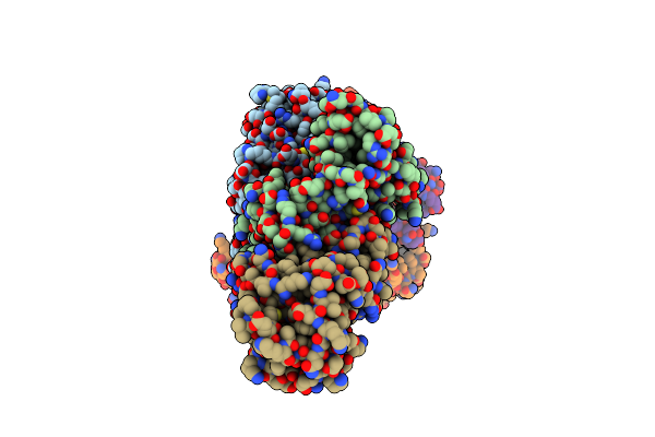
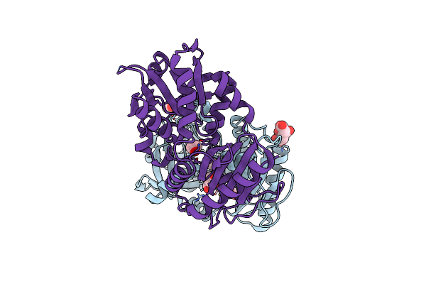
Structure Of Muscarinic Receptor (M2R) In Complex With Beta-Arrestin1 (Local Refine, Cross-Linked)
Organism: Bos taurus, Mus musculus, Homo sapiens
Method: ELECTRON MICROSCOPY Release Date: 2023-12-27 Classification: SIGNALING PROTEIN/IMMUNE SYSTEM |
 |
Organism: Rattus norvegicus, Mus musculus
Method: ELECTRON MICROSCOPY Release Date: 2023-12-27 Classification: SIGNALING PROTEIN/IMMUNE SYSTEM |
 |
Organism: Rattus norvegicus, Mus musculus, Homo sapiens
Method: ELECTRON MICROSCOPY Release Date: 2023-12-27 Classification: SIGNALING PROTEIN/IMMUNE SYSTEM |
 |
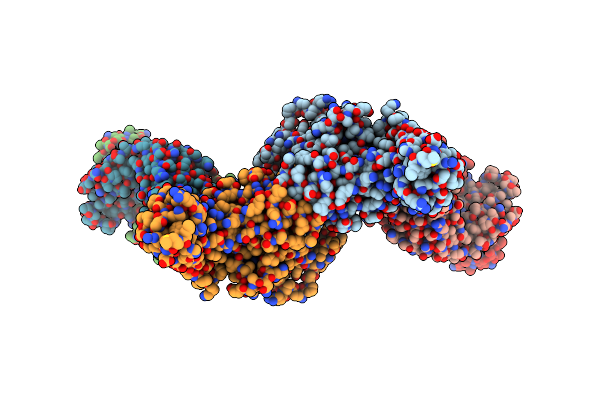
Structure Of Muscarinic Receptor (M2R) In Complex With Beta-Arrestin1 (Local Refine, Non-Cross Linked)
Organism: Bos taurus, Mus musculus, Homo sapiens
Method: ELECTRON MICROSCOPY Release Date: 2023-12-27 Classification: SIGNALING PROTEIN/IMMUNE SYSTEM |
 |
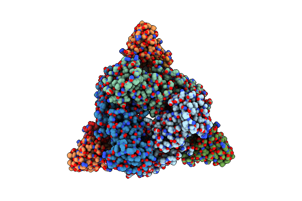
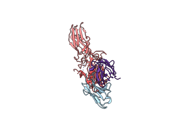
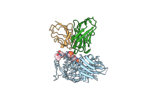
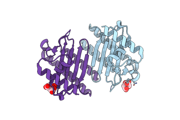
Crystal Structure Of An Antibody Light Chain Tetramer With 3D Domain Swapping
Organism: Homo sapiens
Method: X-RAY DIFFRACTION Resolution:2.00 Å Release Date: 2023-12-27 Classification: IMMUNE SYSTEM |
 |
Organism: Staphylococcus aureus (strain nctc 8325 / ps 47)
Method: X-RAY DIFFRACTION Resolution:2.30 Å Release Date: 2022-11-09 Classification: TOXIN Ligands: MG |
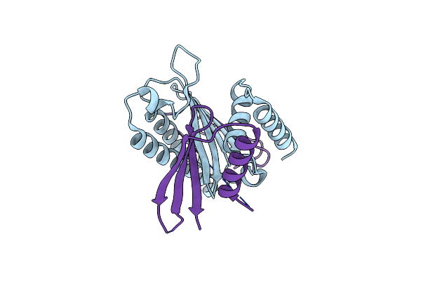 |
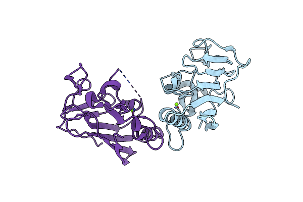
Crystal Structure Of The Nuclease Domain Of Esad In Complex With Esag From Staphylococcus Aureus
Organism: Staphylococcus aureus (strain nctc 8325 / ps 47)
Method: X-RAY DIFFRACTION Resolution:2.59 Å Release Date: 2022-11-09 Classification: TOXIN/ANTITOXIN |
 |
Organism: Staphylococcus aureus (strain nctc 8325 / ps 47)
Method: X-RAY DIFFRACTION Resolution:2.30 Å Release Date: 2022-11-09 Classification: ANTITOXIN Ligands: CIT |
 |
Organism: Homo sapiens
Method: X-RAY DIFFRACTION Resolution:1.79 Å Release Date: 2022-03-02 Classification: SIGNALING PROTEIN, Transferase Ligands: 9ID, SO4 |
 |
Organism: Homo sapiens
Method: X-RAY DIFFRACTION Resolution:1.29 Å Release Date: 2022-02-02 Classification: CYTOSOLIC PROTEIN |
 |
Crystal Structure Of The N-Terminus Of Nonstructural Protein 1 From Sars-Cov-2
Organism: Severe acute respiratory syndrome coronavirus 2
Method: X-RAY DIFFRACTION Resolution:1.25 Å Release Date: 2021-06-09 Classification: VIRAL PROTEIN |
 |
Organism: Mycobacterium tuberculosis (strain atcc 25618 / h37rv)
Method: X-RAY DIFFRACTION Resolution:2.40 Å Release Date: 2020-09-23 Classification: TRANSFERASE Ligands: CIT, EDO |