
Search Count: 44
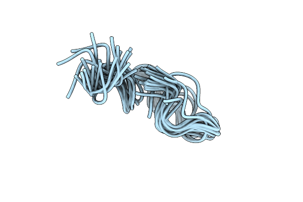 |
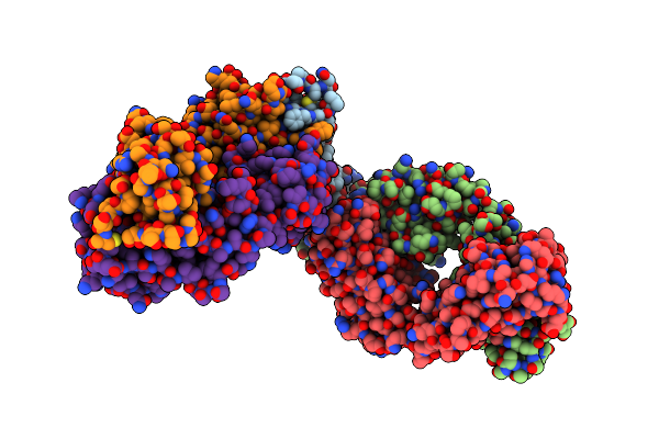
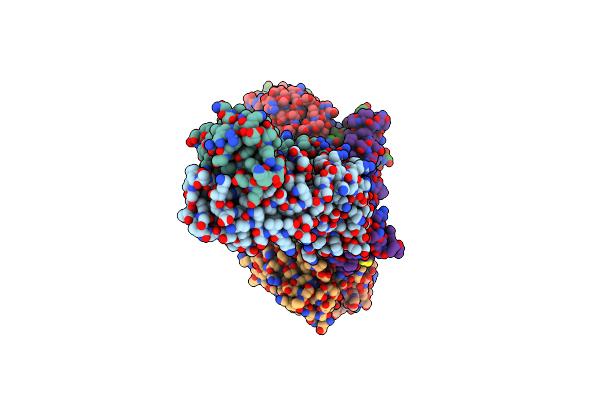
Solution Nmr Structure Of The Bound Antimicrobial Peptide Kg18 To Pseudomonas Aeruginosa Lps Bicelle At Ph 4
Organism: Dengue virus
Method: SOLUTION NMR Release Date: 2025-09-24 Classification: ANTIMICROBIAL PROTEIN |
 |
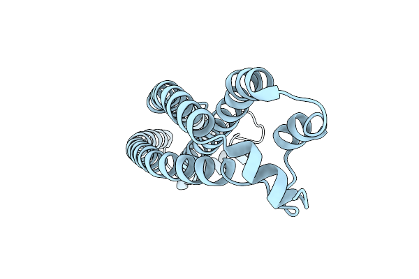
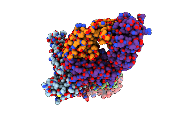
Solution Nmr Structure Of The Bound Antimicrobial Peptide Vr18 To Pseudomonas Aeruginosa Lps Bicelle At Ph 4
Organism: Dengue virus
Method: SOLUTION NMR Release Date: 2025-09-17 Classification: ANTIMICROBIAL PROTEIN |
 |
Organism: Human respiratory syncytial virus a
Method: X-RAY DIFFRACTION Release Date: 2025-08-20 Classification: VIRAL PROTEIN |
 |
Organism: Respiratory syncytial virus
Method: X-RAY DIFFRACTION Release Date: 2025-08-20 Classification: VIRAL PROTEIN |
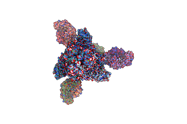 |
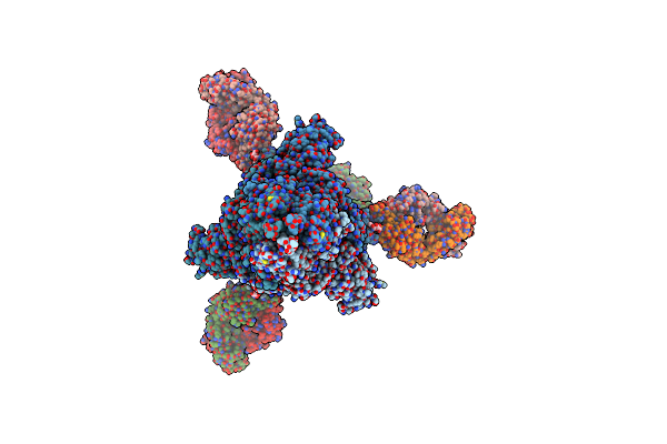
Cryo-Em Structure Of Sars-Cov-2 Omicron Ba.2 S-Trimer In Complex With Fab L4.65 And L5.34
Organism: Severe acute respiratory syndrome coronavirus 2, Homo sapiens
Method: ELECTRON MICROSCOPY Release Date: 2024-01-31 Classification: IMMUNE SYSTEM/VIRAL PROTEIN Ligands: NAG |
 |
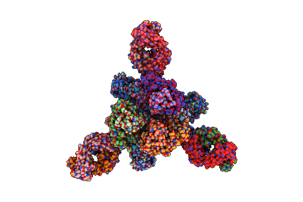
Cryo-Em Structure Of Sars-Cov-2 Omicron Ba.4 S-Trimer In Complex With Fab L4.65 And L5.34
Organism: Severe acute respiratory syndrome coronavirus 2, Homo sapiens
Method: ELECTRON MICROSCOPY Release Date: 2024-01-31 Classification: IMMUNE SYSTEM/VIRAL PROTEIN Ligands: NAG |
 |
Cryo-Em Structure Of Sars-Cov-2 Omicron Prototype S-Trimer In Complex With Fab L4.65 And L5.34
Organism: Severe acute respiratory syndrome coronavirus 2, Homo sapiens
Method: ELECTRON MICROSCOPY Release Date: 2024-01-31 Classification: IMMUNE SYSTEM/VIRAL PROTEIN |
 |
Cryo-Em Structure Of Sars-Cov-2 Omicron Ba.2 Rbd In Complex With Fab L4.65 And L5.34
Organism: Severe acute respiratory syndrome coronavirus 2, Homo sapiens
Method: ELECTRON MICROSCOPY Release Date: 2023-12-20 Classification: IMMUNE SYSTEM/VIRAL PROTEIN Ligands: NAG |
 |
Cryo-Em Structure Of Sars-Cov-2 Omicron Ba.4 Rbd In Complex With Fab L4.65 And L5.34
Organism: Severe acute respiratory syndrome coronavirus 2, Homo sapiens
Method: ELECTRON MICROSCOPY Release Date: 2023-12-20 Classification: IMMUNE SYSTEM/VIRAL PROTEIN Ligands: NAG |
 |
Cryo-Em Structure Of Sars-Cov-2 Omicron Prototype Rbd In Complex With Fab L4.65 And L5.34
Organism: Severe acute respiratory syndrome coronavirus 2, Homo sapiens
Method: ELECTRON MICROSCOPY Release Date: 2023-12-20 Classification: IMMUNE SYSTEM/VIRAL PROTEIN |
 |
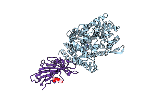
Organism: Plasmodium vivax
Method: X-RAY DIFFRACTION Resolution:1.45 Å Release Date: 2023-07-26 Classification: CELL INVASION |
 |
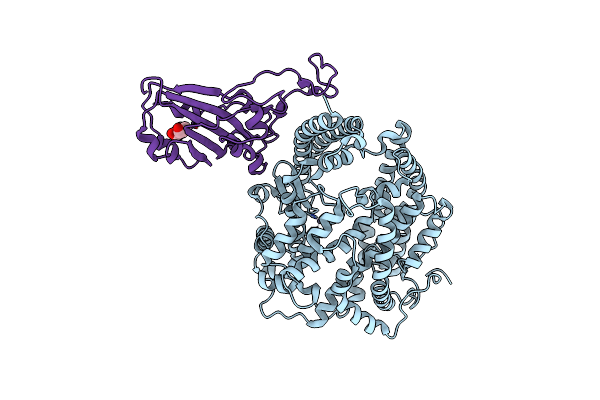
Crystal Structure Of Fada2 (Rv0243) From The Fatty Acid Metabolic Pathway Of Mycobacterium Tuberculosis
Organism: Mycobacterium tuberculosis h37rv
Method: X-RAY DIFFRACTION Resolution:2.90 Å Release Date: 2023-04-26 Classification: TRANSFERASE Ligands: SO4 |
 |
Organism: Severe acute respiratory syndrome coronavirus 2, Vicugna pacos
Method: X-RAY DIFFRACTION Resolution:2.50 Å Release Date: 2022-12-21 Classification: VIRAL PROTEIN |
 |
Organism: Severe acute respiratory syndrome coronavirus 2, Vicugna pacos
Method: X-RAY DIFFRACTION Resolution:2.69 Å Release Date: 2022-12-21 Classification: VIRAL PROTEIN Ligands: NAG |
 |
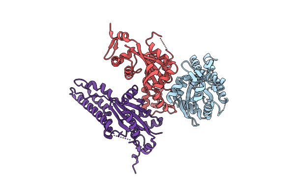
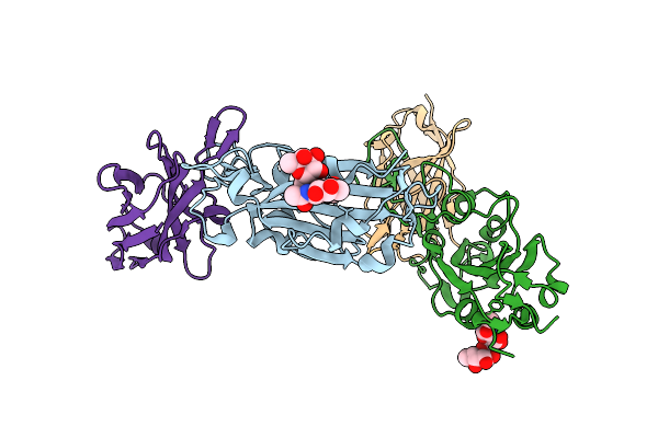
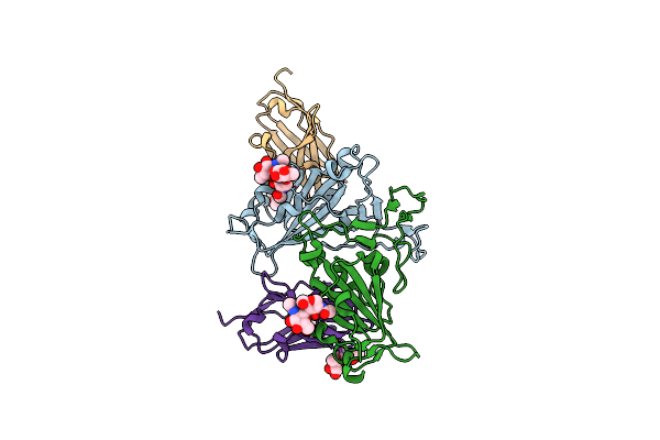
Crystal Structure Of Malaria Transmission-Blocking Antigen Pfs48/45-6C Variant In Complex With Human Antibodies Rupa-117 And Rupa-47
Organism: Homo sapiens, Plasmodium falciparum
Method: X-RAY DIFFRACTION Resolution:2.18 Å Release Date: 2022-08-10 Classification: IMMUNE SYSTEM Ligands: NAG |
 |
Organism: Homo sapiens, Severe acute respiratory syndrome coronavirus 2
Method: ELECTRON MICROSCOPY Release Date: 2022-07-06 Classification: VIRAL PROTEIN/IMMUNE SYSTEM |
 |
Organism: Severe acute respiratory syndrome coronavirus 2, Homo sapiens
Method: ELECTRON MICROSCOPY Release Date: 2022-06-22 Classification: VIRAL PROTEIN/IMMUNE SYSTEM Ligands: NAG, MES |
 |
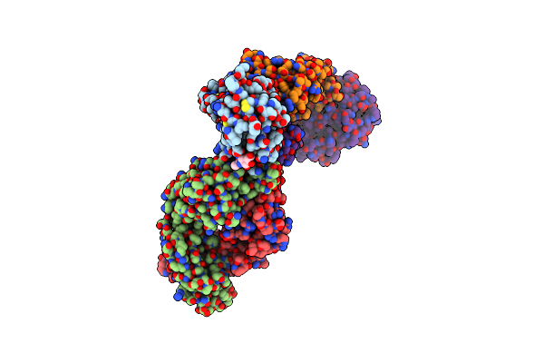
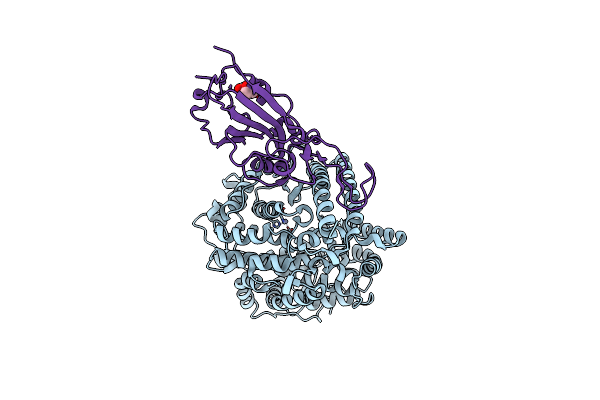
Structure Of Bat Coronavirus Ratg13 Spike Receptor-Binding Domain Complexed With Its Receptor Equine Ace2
Organism: Equus caballus, Bat coronavirus ratg13
Method: X-RAY DIFFRACTION Resolution:2.60 Å Release Date: 2022-06-08 Classification: VIRAL PROTEIN/INHIBITOR Ligands: ZN, NAG |
 |
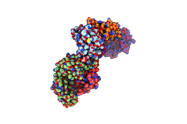
Structure Of Sars-Cov-2 Spike Receptor-Binding Domain Complexed With Its Receptor Equine Ace2
Organism: Equus caballus, Severe acute respiratory syndrome coronavirus 2
Method: X-RAY DIFFRACTION Resolution:2.56 Å Release Date: 2022-06-08 Classification: VIRAL PROTEIN/INHIBITOR Ligands: ZN, NAG |
 |
The Crystal Structure Of Sars-Cov-2 Omicron Ba.1 Variant Rbd In Complex With Equine Ace2
Organism: Equus caballus, Severe acute respiratory syndrome coronavirus 2
Method: X-RAY DIFFRACTION Resolution:2.85 Å Release Date: 2022-06-08 Classification: VIRAL PROTEIN Ligands: ZN, BR, NAG |