
Search Count: 127
 |
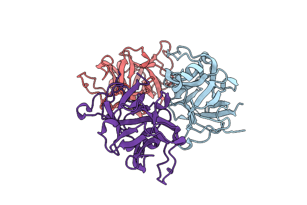
Organism: Homo sapiens
Method: X-RAY DIFFRACTION Release Date: 2025-12-17 Classification: IMMUNE SYSTEM |
 |
Organism: Saccharomyces cerevisiae
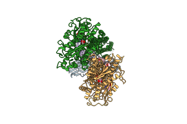
Method: ELECTRON MICROSCOPY Release Date: 2025-10-15 Classification: TRANSFERASE Ligands: CPL |
 |
Organism: Saccharomyces cerevisiae
Method: ELECTRON MICROSCOPY Release Date: 2025-10-15 Classification: MEMBRANE PROTEIN Ligands: NNM |
 |
Organism: Saccharomyces cerevisiae
Method: ELECTRON MICROSCOPY Release Date: 2025-10-15 Classification: MEMBRANE PROTEIN Ligands: CPL, MAN |
 |
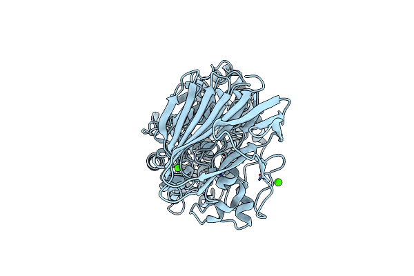
Crystal Structure Of The Mir Domain Of The S. Cerevisiae Mannosyltransferase Pmt4
Organism: Saccharomyces cerevisiae
Method: X-RAY DIFFRACTION Release Date: 2025-10-15 Classification: MEMBRANE PROTEIN |
 |
Crystal Structure Of The Mir Domain Of The S. Cerevisiae Mannosyltransferase Pmt4 In Complex With Ccw5
Organism: Saccharomyces cerevisiae
Method: X-RAY DIFFRACTION Release Date: 2025-10-15 Classification: MEMBRANE PROTEIN Ligands: PEG, GOL |
 |
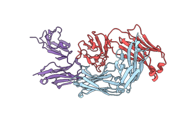
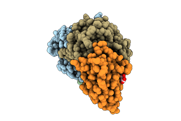
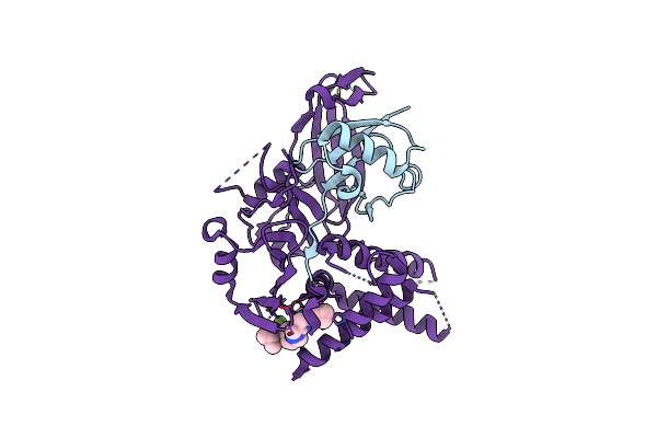
Organism: Homo sapiens, Human coronavirus hku1 (isolate n1)
Method: X-RAY DIFFRACTION Release Date: 2025-07-30 Classification: VIRAL PROTEIN Ligands: CA, NAG |
 |
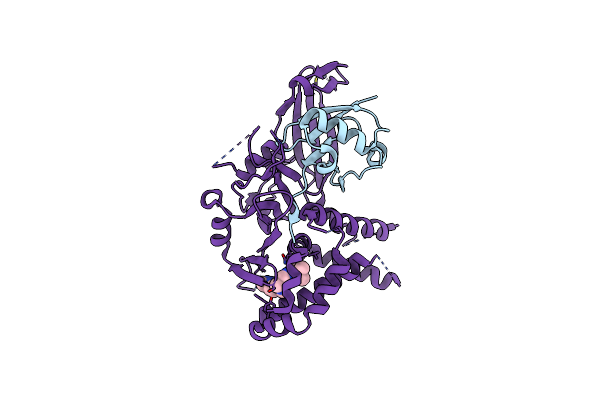
Organism: Human coronavirus hku1 (isolate n1), Homo sapiens
Method: X-RAY DIFFRACTION Release Date: 2025-07-30 Classification: ANTIVIRAL PROTEIN Ligands: NAG, GOL |
 |
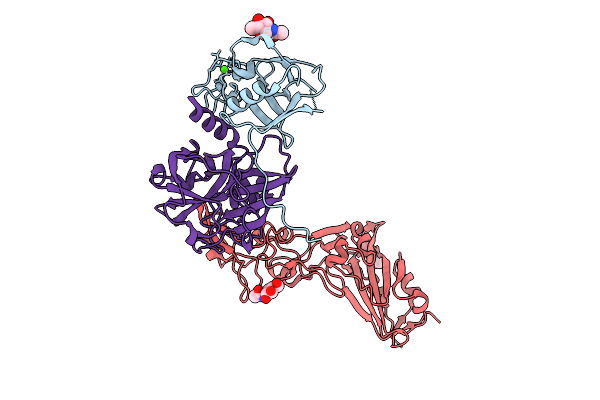
Organism: Homo sapiens, Vicugna pacos
Method: X-RAY DIFFRACTION Release Date: 2025-07-30 Classification: ANTIVIRAL PROTEIN Ligands: CA, NAG, GOL, MLI |
 |
Organism: Homo sapiens
Method: X-RAY DIFFRACTION Release Date: 2025-07-30 Classification: ANTIVIRAL PROTEIN Ligands: MES, GOL, CA |
 |
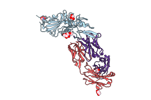
Organism: Homo sapiens, Vicugna pacos
Method: X-RAY DIFFRACTION Release Date: 2025-07-30 Classification: IMMUNE SYSTEM Ligands: CA, NAG, GOL |
 |
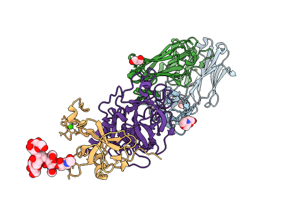
Multibody Refinement Of Dimeric Darpin And Its Bound Gfp On A Symmetric Scaffold
Organism: Synthetic construct, Aequorea victoria
Method: ELECTRON MICROSCOPY Release Date: 2025-06-04 Classification: BIOSYNTHETIC PROTEIN |
 |
24-Mer Darpin-Apoferritin Scaffold In Complex With The Maltose Binding Protein
Organism: Synthetic construct, Homo sapiens, Escherichia coli
Method: ELECTRON MICROSCOPY Release Date: 2025-06-04 Classification: BIOSYNTHETIC PROTEIN |
 |
Organism: Homo sapiens, Aequorea victoria
Method: ELECTRON MICROSCOPY Release Date: 2025-06-04 Classification: METAL BINDING PROTEIN/LUMINESCENT PROTEIN |
 |
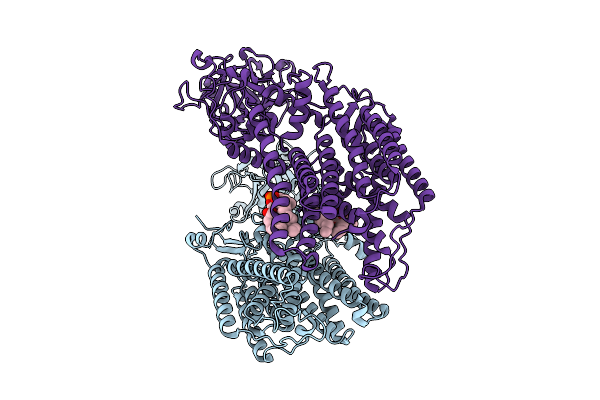
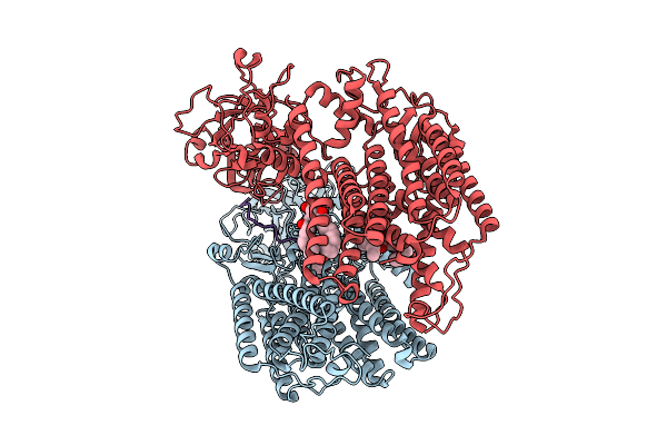
Organism: Homo sapiens, Mus musculus
Method: ELECTRON MICROSCOPY Release Date: 2025-03-05 Classification: MEMBRANE PROTEIN Ligands: GLU, CLR |
 |
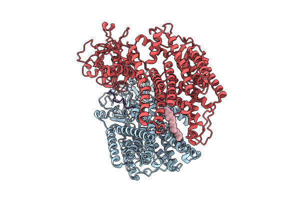
Organism: Homo sapiens, Mus musculus
Method: ELECTRON MICROSCOPY Release Date: 2025-03-05 Classification: MEMBRANE PROTEIN Ligands: GLU, CLR |
 |
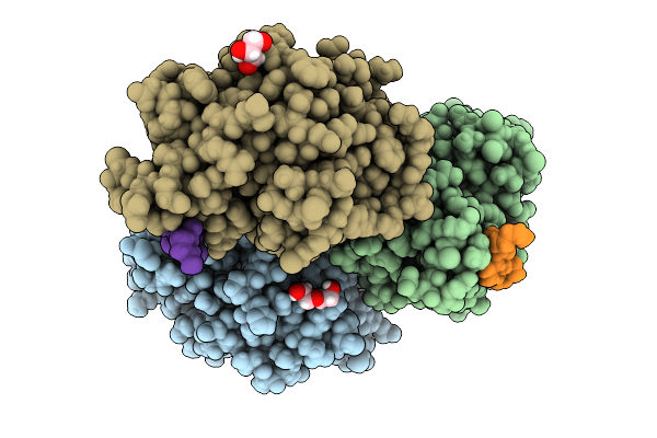
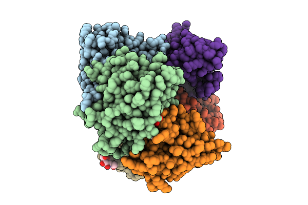
Organism: Spirosoma fluviale
Method: X-RAY DIFFRACTION Release Date: 2024-09-04 Classification: LYASE Ligands: MN, CA |
 |
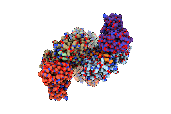
Usp1 Bound To Ksq-4279 And Ubiquitin Conjugated To Fancd2 (Focused Refinement)
Organism: Homo sapiens
Method: ELECTRON MICROSCOPY Release Date: 2024-09-04 Classification: HYDROLASE Ligands: ZN, A1IB8 |
 |
Usp1 Bound To Ml323 And Ubiquitin Conjugated To Fancd2 (Ordered Subset, Focused Refinement)
Organism: Homo sapiens
Method: ELECTRON MICROSCOPY Release Date: 2024-09-04 Classification: HYDROLASE Ligands: ZN, JDA |
 |
Organism: Drosophila melanogaster, Homo sapiens
Method: SOLUTION NMR Release Date: 2024-08-14 Classification: LIGASE Ligands: ZN |