
Search Count: 60
 |
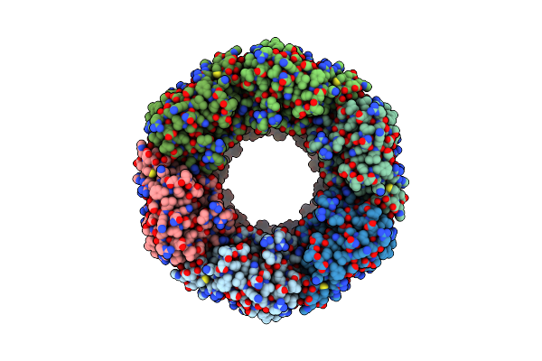
Organism: Triticum aestivum
Method: ELECTRON MICROSCOPY Resolution:3.33 Å Release Date: 2025-12-10 Classification: PLANT PROTEIN |
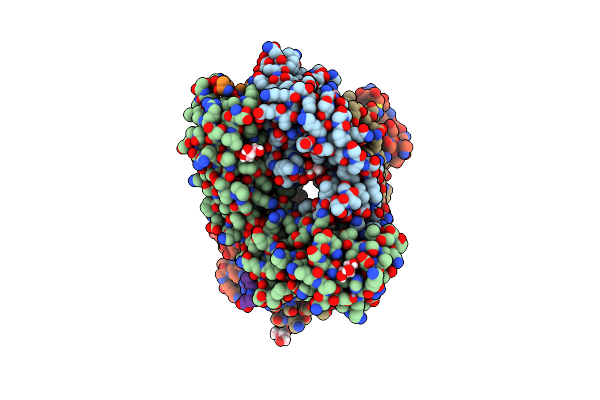 |
Organism: Synthetic construct, Homo sapiens
Method: X-RAY DIFFRACTION Resolution:2.00 Å Release Date: 2024-07-31 Classification: IMMUNE SYSTEM/PEPTIDE BINDING PROTEIN Ligands: GOL, ZN, PEG |
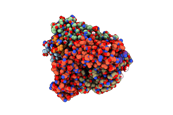 |
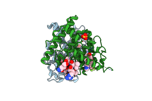
Organism: Homo sapiens
Method: X-RAY DIFFRACTION Resolution:1.90 Å Release Date: 2024-06-19 Classification: PEPTIDE BINDING PROTEIN Ligands: SO4, ZN, GOL, ACT, PEG |
 |
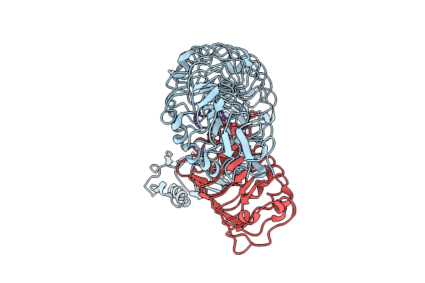
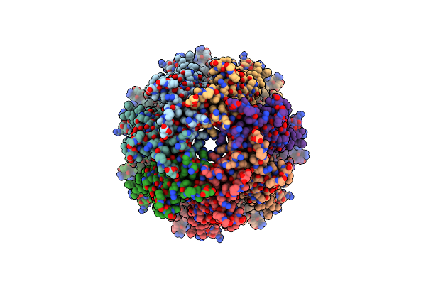
Organism: Alkalihalophilus pseudofirmus of4
Method: X-RAY DIFFRACTION Resolution:3.20 Å Release Date: 2023-12-13 Classification: HYDROLASE Ligands: CA |
 |
Phage Display Derived Serum Albumin Binding Knob Domain Engineered Within A Novel Vh Framework 3 Bispecific Antibody Format
Organism: Cricetulus griseus
Method: X-RAY DIFFRACTION Resolution:2.00 Å Release Date: 2023-06-07 Classification: IMMUNE SYSTEM Ligands: CL |
 |
Phage Display Derived Serum Albumin Binding Knob Domain Engineered Within A Novel Vh Framework 3 Bispecific Antibody Format
Organism: Cricetulus griseus
Method: X-RAY DIFFRACTION Resolution:1.61 Å Release Date: 2023-06-07 Classification: IMMUNE SYSTEM |
 |
Estrogen Receptor Alpha Ligand Binding Domain Y537S Mutant In Complex With An Inhibitor 30O And Grip Peptide
Organism: Homo sapiens
Method: X-RAY DIFFRACTION Resolution:2.22 Å Release Date: 2023-04-26 Classification: TRANSCRIPTION/INHIBITOR Ligands: PEG, IC7, SO4 |
 |
Estrogen Receptor Alpha Ligand Binding Domain Y537S Mutant In Complex With An Inhibitor 30A And Grip Peptide
Organism: Homo sapiens
Method: X-RAY DIFFRACTION Resolution:2.14 Å Release Date: 2023-04-26 Classification: TRANSCRIPTION/INHIBITOR Ligands: IAT, PEG |
 |
Organism: Homo sapiens
Method: ELECTRON MICROSCOPY Release Date: 2023-03-15 Classification: MEMBRANE PROTEIN |
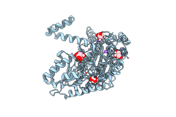 |
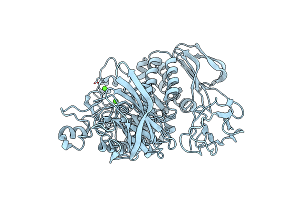
Organism: Aspergillus fumigatus
Method: X-RAY DIFFRACTION Resolution:1.56 Å Release Date: 2022-08-17 Classification: ISOMERASE Ligands: GOL, FLC, CL, NA |
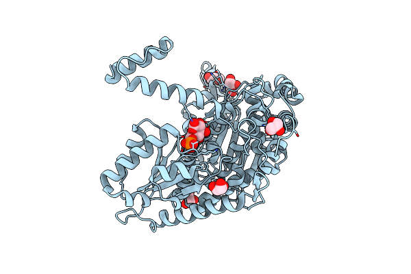 |
Phosphoglucose Isomerase Of Aspergillus Fumigatus In Complexed With Glucose-6-Phosphate
Organism: Neosartorya fumigata
Method: X-RAY DIFFRACTION Resolution:1.78 Å Release Date: 2022-07-13 Classification: ISOMERASE Ligands: CL, GOL, NA, BG6 |
 |
Organism: Arabidopsis thaliana
Method: ELECTRON MICROSCOPY Release Date: 2021-09-22 Classification: PLANT PROTEIN |
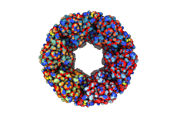 |
Organism: Arabidopsis thaliana
Method: ELECTRON MICROSCOPY Release Date: 2021-09-22 Classification: PLANT PROTEIN |
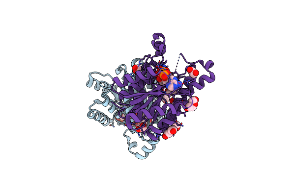 |
Promiscuous Reductase Lugoii Catalyzes Keto-Reduction At C1 During Lugdunomycin Biosynthesis
Organism: Streptomyces sp. ql37
Method: X-RAY DIFFRACTION Resolution:1.08 Å Release Date: 2020-09-16 Classification: ANTIBIOTIC Ligands: NDP, EDO, PEG |
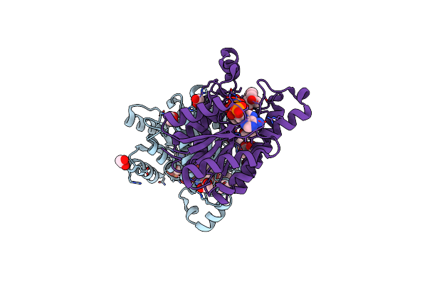 |
Promiscuous Reductase Lugoii Catalyzes Keto-Reduction At C1 During Lugdunomycin Biosynthesis
Organism: Streptomyces sp. ql37
Method: X-RAY DIFFRACTION Resolution:1.08 Å Release Date: 2020-09-16 Classification: ANTIBIOTIC Ligands: EDO, NAP, P7K |
 |
Promiscuous Reductase Lugoii Catalyzes Keto-Reduction At C1 During Lugdunomycin Biosynthesis
Organism: Streptomyces sp. ql37
Method: X-RAY DIFFRACTION Resolution:1.57 Å Release Date: 2020-09-16 Classification: ANTIBIOTIC Ligands: NAP, P7Q, EDO |
 |
Promiscuous Reductase Lugoii Catalyzes Keto-Reduction At C1 During Lugdunomycin Biosynthesis
Organism: Streptomyces sp. ql37
Method: X-RAY DIFFRACTION Resolution:2.08 Å Release Date: 2020-09-16 Classification: ANTIBIOTIC Ligands: EDO, PEG, SO4 |
 |
Organism: Mycobacterium tuberculosis h37rv
Method: X-RAY DIFFRACTION Resolution:2.20 Å Release Date: 2020-08-05 Classification: TOXIN |
 |
Organism: Mycobacterium tuberculosis h37rv
Method: X-RAY DIFFRACTION Resolution:2.00 Å Release Date: 2020-08-05 Classification: TOXIN |
 |
Organism: Mycobacterium tuberculosis
Method: X-RAY DIFFRACTION Resolution:2.30 Å Release Date: 2020-08-05 Classification: TOXIN |