
Search Count: 45
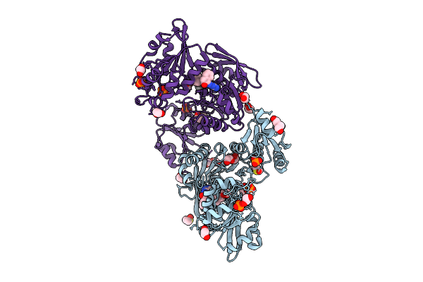 |
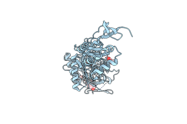
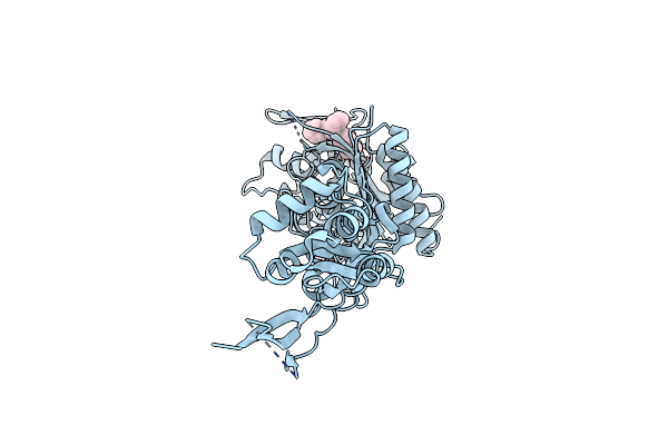
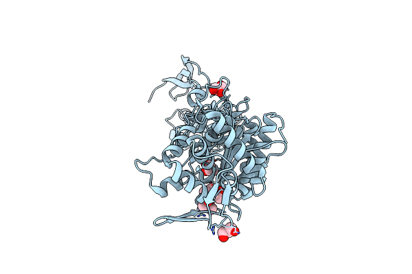
Crystal Structure Of Udp-N-Acetylmuramate-L-Alanine Ligase (Murc) From Pseudomonas Aeruginosa In Complex With Compound Osa_001176 (Wyh78)
Organism: Pseudomonas aeruginosa
Method: X-RAY DIFFRACTION Release Date: 2025-09-17 Classification: BIOSYNTHETIC PROTEIN Ligands: WIR, PO4, UNX, EDO, PEG, DMS |
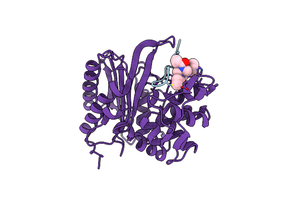 |
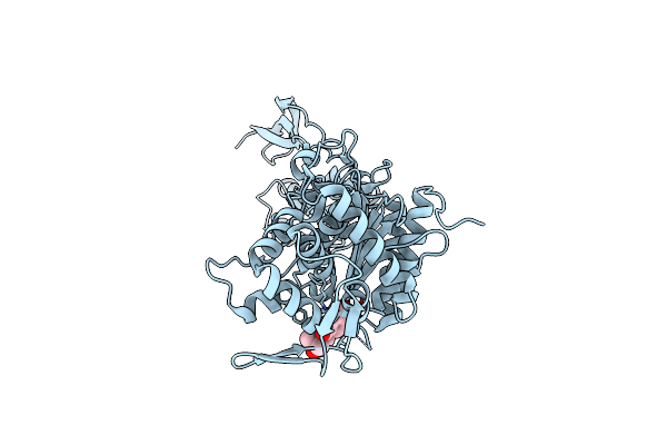
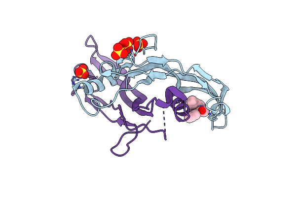
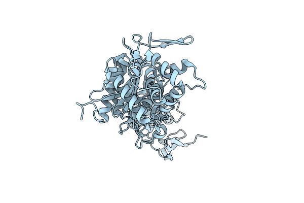
The Structure Of E. Coli Penicillin Binding Protein 3 (Pbp3) In Complex With A Bicyclic Peptide Inhibitor
Organism: Escherichia coli, Synthetic construct
Method: X-RAY DIFFRACTION Release Date: 2024-04-03 Classification: HYDROLASE Ligands: 29N |
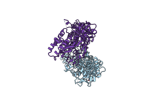 |
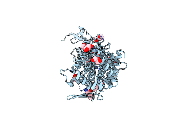
Organism: Acinetobacter baumannii
Method: X-RAY DIFFRACTION Resolution:2.65 Å Release Date: 2023-02-22 Classification: HYDROLASE Ligands: ZN |
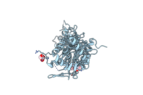 |
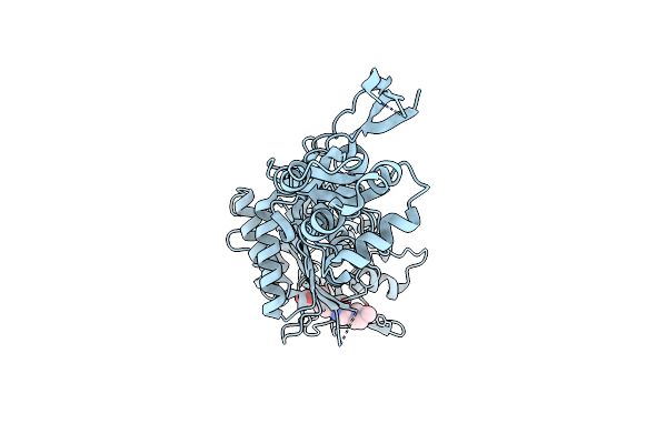
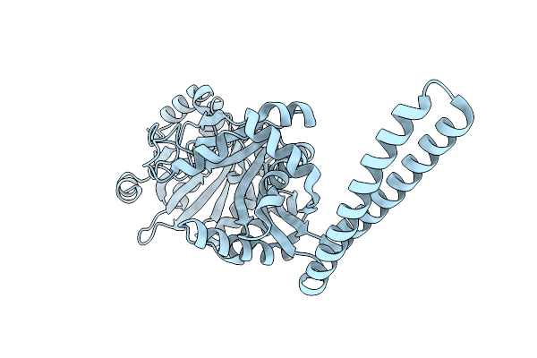
Structure Of P. Aeruginosa Pbp3 In Complex With A Phenyl Boronic Acid (Compound 1)
Organism: Pseudomonas aeruginosa pao1
Method: X-RAY DIFFRACTION Resolution:1.58 Å Release Date: 2021-08-11 Classification: HYDROLASE Ligands: GOL, RY2, DMS |
 |
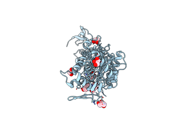
Structure Of P. Aeruginosa Pbp3 In Complex With An Aryl Boronic Acid (Compound 2)
Organism: Pseudomonas aeruginosa pao1
Method: X-RAY DIFFRACTION Resolution:1.59 Å Release Date: 2021-08-11 Classification: HYDROLASE Ligands: RXW, DMS, GOL |
 |
Structure Of P. Aeruginosa Pbp3 In Complex With A Benzoxaborole (Compound 3)
Organism: Pseudomonas aeruginosa
Method: X-RAY DIFFRACTION Resolution:1.44 Å Release Date: 2021-08-11 Classification: HYDROLASE Ligands: GOL, RZZ |
 |
Structure Of P. Aeruginosa Pbp3 In Complex With A Benzoxaborole (Compound 4)
Organism: Pseudomonas aeruginosa
Method: X-RAY DIFFRACTION Resolution:1.80 Å Release Date: 2021-08-11 Classification: HYDROLASE Ligands: S1B |
 |
Structure Of P. Aeruginosa Pbp3 In Complex With A Benzoxaborole (Compound 7)
Organism: Pseudomonas aeruginosa
Method: X-RAY DIFFRACTION Resolution:2.17 Å Release Date: 2021-08-11 Classification: HYDROLASE Ligands: S0Z |
 |
Structure Of P. Aeruginosa Pbp3 In Complex With A Benzoxaborole (Compound 12)
Organism: Pseudomonas aeruginosa
Method: X-RAY DIFFRACTION Resolution:1.36 Å Release Date: 2021-08-11 Classification: HYDROLASE Ligands: DMS, PEG, S08, GOL |
 |
Structure Of P. Aeruginosa Pbp3 In Complex With A Benzoxaborole (Compound 13)
Organism: Pseudomonas aeruginosa
Method: X-RAY DIFFRACTION Resolution:1.79 Å Release Date: 2021-08-11 Classification: HYDROLASE Ligands: S0Q |
 |
Structure Of P. Aeruginosa Pbp3 In Complex With A Benzoxaborole (Compound 14)
Organism: Pseudomonas aeruginosa
Method: X-RAY DIFFRACTION Resolution:2.14 Å Release Date: 2021-08-11 Classification: HYDROLASE Ligands: GOL, S1E |
 |
Structure Of P. Aeruginosa Pbp3 In Complex With A Benzoxaborole (Compound 15)
Organism: Pseudomonas aeruginosa
Method: X-RAY DIFFRACTION Resolution:1.91 Å Release Date: 2021-08-11 Classification: HYDROLASE Ligands: S1K |
 |
Organism: Pseudomonas aeruginosa
Method: X-RAY DIFFRACTION Resolution:2.01 Å Release Date: 2021-08-11 Classification: HYDROLASE Ligands: GOL, 4D6 |
 |
Organism: Homo sapiens
Method: X-RAY DIFFRACTION Resolution:2.04 Å Release Date: 2021-02-03 Classification: SIGNALING PROTEIN Ligands: SO4, OCK |
 |
Organism: Homo sapiens
Method: X-RAY DIFFRACTION Resolution:2.03 Å Release Date: 2021-01-13 Classification: SIGNALING PROTEIN Ligands: ODQ, SO4 |
 |
Organism: Staphylococcus aureus
Method: X-RAY DIFFRACTION Resolution:1.62 Å Release Date: 2020-09-09 Classification: ANTIMICROBIAL PROTEIN |
 |
Structure Of Pseudomonas Aeruginosa Penicillin-Binding Protein 3 (Pbp3) In Complex With Compound 6
Organism: Pseudomonas aeruginosa (strain atcc 15692 / dsm 22644 / cip 104116 / jcm 14847 / lmg 12228 / 1c / prs 101 / pao1)
Method: X-RAY DIFFRACTION Resolution:1.55 Å Release Date: 2020-06-24 Classification: HYDROLASE Ligands: GOL, ODZ |
 |
Structure Of Pseudomonas Aeruginosa Penicillin-Binding Protein 3 (Pbp3) In Complex With Compound 1
Organism: Pseudomonas aeruginosa (strain atcc 15692 / dsm 22644 / cip 104116 / jcm 14847 / lmg 12228 / 1c / prs 101 / pao1)
Method: X-RAY DIFFRACTION Resolution:1.70 Å Release Date: 2020-06-24 Classification: HYDROLASE Ligands: OEE, GOL |
 |
Structure Of Pseudomonas Aeruginosa Penicillin-Binding Protein 3 (Pbp3) In Complex With Piperacillin
Organism: Pseudomonas aeruginosa (strain atcc 15692 / dsm 22644 / cip 104116 / jcm 14847 / lmg 12228 / 1c / prs 101 / pao1)
Method: X-RAY DIFFRACTION Resolution:1.59 Å Release Date: 2020-02-19 Classification: HYDROLASE Ligands: JPP |
 |
Apo Structure Of R504C Mutant Of Pseudomonas Aeruginosa Penicillin-Binding Protein 3 (Pbp3)
Organism: Pseudomonas aeruginosa (strain atcc 15692 / dsm 22644 / cip 104116 / jcm 14847 / lmg 12228 / 1c / prs 101 / pao1)
Method: X-RAY DIFFRACTION Resolution:2.20 Å Release Date: 2020-02-19 Classification: HYDROLASE |