
Search Count: 16
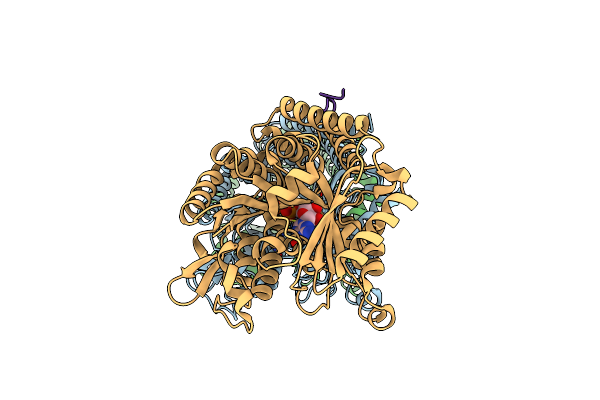 |
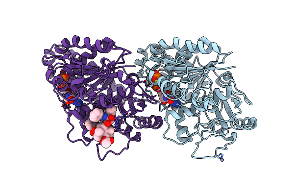
Model Of Synthetic Tau (Four Tandem Repeats Of First Repeat Sequence) Bound To The Microtubule
Organism: Homo sapiens, Sus scrofa
Method: ELECTRON MICROSCOPY Release Date: 2018-05-23 Classification: STRUCTURAL PROTEIN Ligands: GTP, MG, GDP |
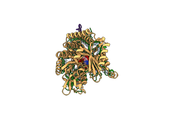 |
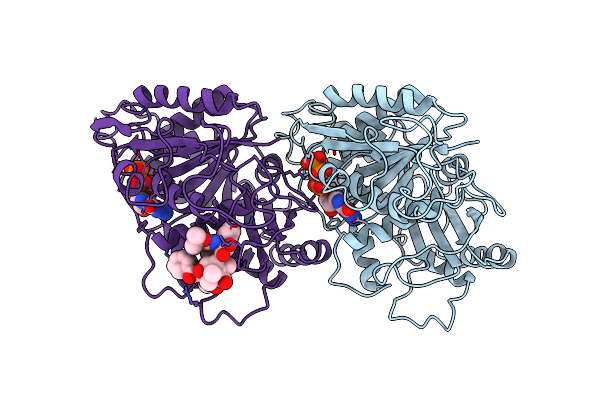
Organism: Homo sapiens, Sus scrofa
Method: ELECTRON MICROSCOPY Release Date: 2018-05-23 Classification: STRUCTURAL PROTEIN Ligands: GDP, GTP, MG |
 |
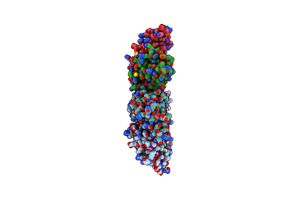
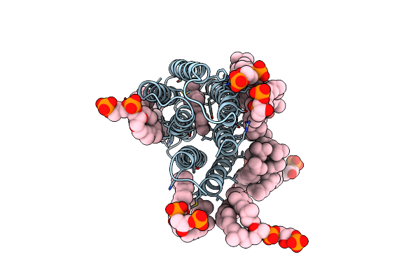
Near-Atomic Resolution Cryo-Em Reconstruction Of Peloruside-Stabilized Microtubule
Organism: Sus scrofa
Method: ELECTRON MICROSCOPY Release Date: 2017-02-01 Classification: STRUCTURAL PROTEIN Ligands: GTP, MG, GDP, POU |
 |
Near-Atomic Resolution Cryo-Em Reconstruction Of Doubly Bound Taxol- And Peloruside-Stabilized Microtubule
Organism: Sus scrofa
Method: ELECTRON MICROSCOPY Release Date: 2017-02-01 Classification: STRUCTURAL PROTEIN Ligands: GTP, MG, GDP, TA1, POU |
 |
Organism: Sus scrofa
Method: ELECTRON MICROSCOPY Release Date: 2017-02-01 Classification: STRUCTURAL PROTEIN Ligands: GTP, MG, GDP, TA1 |
 |
Organism: Sus scrofa
Method: ELECTRON MICROSCOPY Release Date: 2017-02-01 Classification: STRUCTURAL PROTEIN Ligands: GTP, MG, GDP, ZPN |
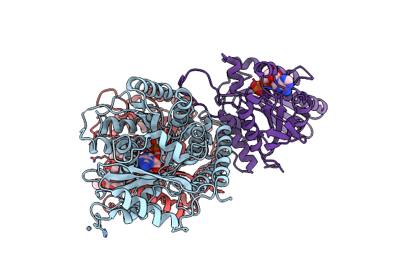 |
Human Monomeric Kinesin (1Bg2) And Bovine Tubulin (1Jff) Docked Into The 9-Angstrom Cryo-Em Map Of Nucleotide-Free Kinesin Complexed To The Microtubule
Organism: Homo sapiens, Bos taurus
Method: ELECTRON MICROSCOPY Release Date: 2008-07-08 Classification: TRANSPORT PROTEIN Ligands: MG, ADP, ZN, GTP, GDP, TA1 |
 |
Organism: Caenorhabditis elegans
Method: X-RAY DIFFRACTION Resolution:2.64 Å Release Date: 2006-02-07 Classification: TRANSFERASE |
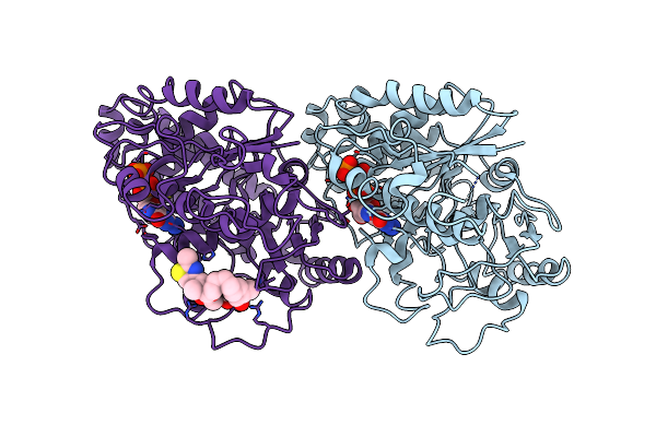 |
The Binding Mode Of Epothilone A On A,B-Tubulin By Electron Crystallography
Organism: Bos taurus
Method: ELECTRON CRYSTALLOGRAPHY Resolution:2.89 Å Release Date: 2004-09-14 Classification: CELL CYCLE, STRUCTURAL PROTEIN Ligands: GTP, GDP, EP |
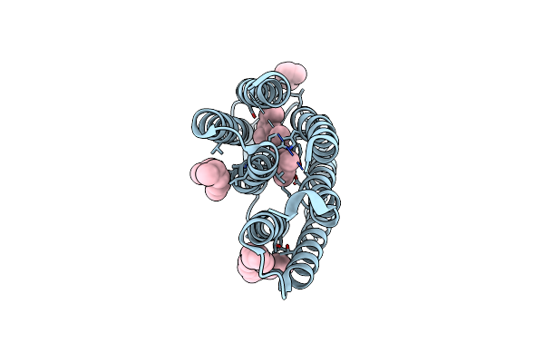 |
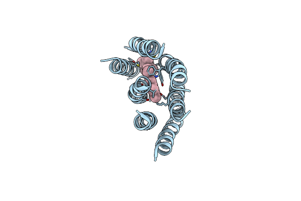
Organism: Halobacterium salinarum
Method: X-RAY DIFFRACTION Resolution:2.00 Å Release Date: 2001-12-05 Classification: PROTON TRANSPORT Ligands: RET, LI1 |
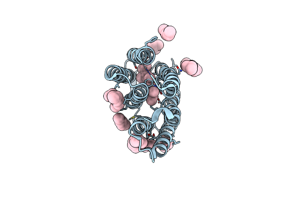 |
Organism: Halobacterium salinarum
Method: X-RAY DIFFRACTION Resolution:1.81 Å Release Date: 2001-12-05 Classification: PROTON TRANSPORT Ligands: RET, LI1 |
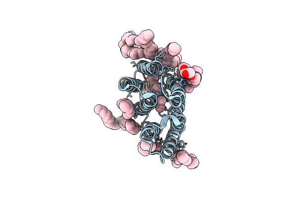 |
Organism: Halobacterium salinarum
Method: X-RAY DIFFRACTION Resolution:1.65 Å Release Date: 2001-12-05 Classification: PROTON TRANSPORT Ligands: RET, LI1 |
 |
Refined Structure Of Alpha-Beta Tubulin From Zinc-Induced Sheets Stabilized With Taxol
Organism: Bos taurus
Method: ELECTRON CRYSTALLOGRAPHY Resolution:3.50 Å Release Date: 2001-09-19 Classification: STRUCTURAL PROTEIN Ligands: ZN, MG, GTP, GDP, TA1 |
 |
Organism: Sus scrofa
Method: ELECTRON CRYSTALLOGRAPHY Resolution:3.70 Å Release Date: 1998-10-07 Classification: MICROTUBULES Ligands: GTP, GDP, TXL |
 |
Organism: Halobacterium salinarum
Method: ELECTRON CRYSTALLOGRAPHY Resolution:3.50 Å Release Date: 1996-06-10 Classification: PHOTORECEPTOR Ligands: DPG, RET |
 |
Model For The Structure Of Bacteriorhodopsin Based On High-Resolution Electron Cryo-Microscopy
Organism: Halobacterium salinarum
Method: ELECTRON CRYSTALLOGRAPHY Resolution:3.50 Å Release Date: 1991-04-15 Classification: PHOTORECEPTOR Ligands: RET |