
Search Count: 14
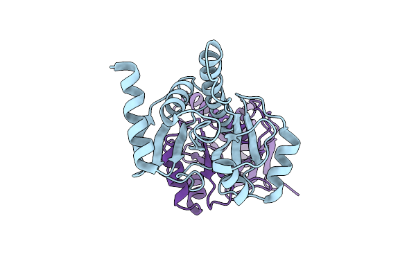 |
Organism: Rubella virus
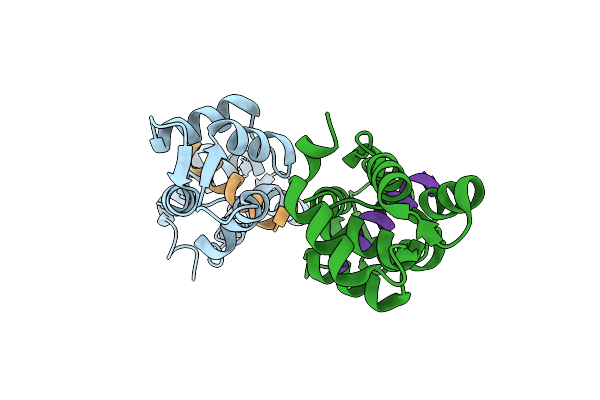
Method: X-RAY DIFFRACTION Resolution:1.72 Å Release Date: 2024-01-17 Classification: HYDROLASE |
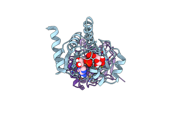 |
Organism: Rubella virus
Method: X-RAY DIFFRACTION Resolution:1.59 Å Release Date: 2024-01-17 Classification: HYDROLASE Ligands: APR |
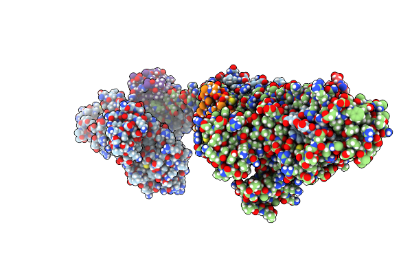 |
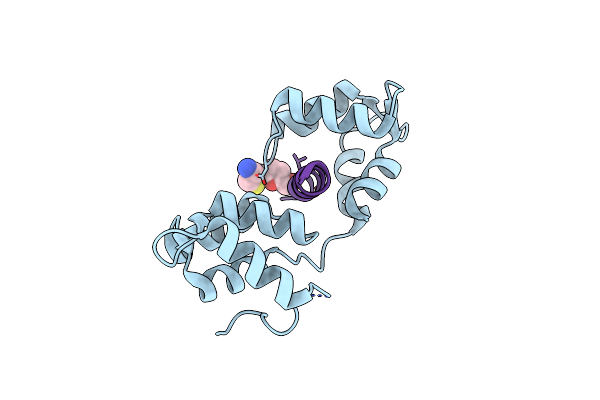
Crystal Structure Of The Kap1 Tripartite Motif In Complex With The Znf93 Krab Domain
Organism: Homo sapiens
Method: X-RAY DIFFRACTION Resolution:2.80 Å Release Date: 2022-11-02 Classification: TRANSCRIPTION Ligands: ZN |
 |
Organism: Homo sapiens
Method: X-RAY DIFFRACTION Resolution:2.03 Å Release Date: 2020-09-16 Classification: GENE REGULATION Ligands: GOL |
 |
Organism: Homo sapiens
Method: X-RAY DIFFRACTION Resolution:2.51 Å Release Date: 2020-08-05 Classification: GENE REGULATION Ligands: SO4 |
 |
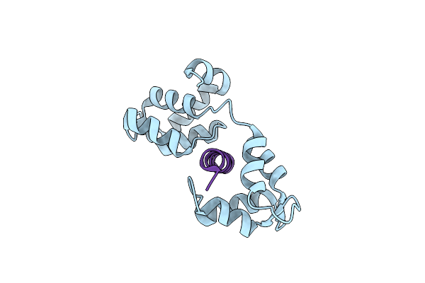
Organism: Homo sapiens
Method: X-RAY DIFFRACTION Resolution:1.81 Å Release Date: 2018-02-14 Classification: NUCLEAR PROTEIN Ligands: ZN, ANP, MG |
 |
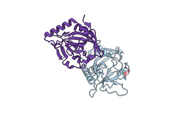
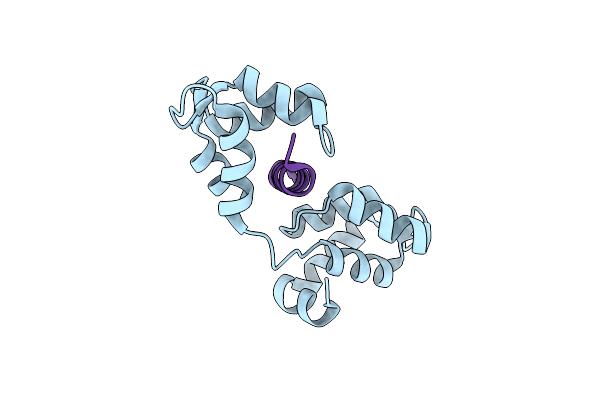
Crystal Structure Of Human Morc2 (Residues 1-603) With Spinal Muscular Atrophy Mutation T424R
Organism: Homo sapiens
Method: X-RAY DIFFRACTION Resolution:2.57 Å Release Date: 2018-02-14 Classification: NUCLEAR PROTEIN Ligands: ZN, ANP, MG |
 |
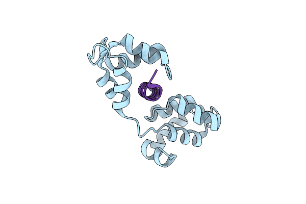
Crystal Structure Of Human Morc2 (Residues 1-603) With Spinal Muscular Atrophy Mutation S87L
Organism: Homo sapiens
Method: X-RAY DIFFRACTION Resolution:2.02 Å Release Date: 2018-02-14 Classification: NUCLEAR PROTEIN Ligands: ZN, ATP, MG |
 |
Solution Structure Of Tpsb4 N-Terminal Potra Domain From Pseudomonas Aeruginosa
Organism: Pseudomonas aeruginosa
Method: SOLUTION NMR Release Date: 2014-12-24 Classification: PROTEIN TRANSPORT |
 |
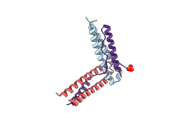
Crystal Structure Of Mtip From Plasmodium Falciparum In Complex With A Peptide-Fragment Chimera
Organism: Plasmodium falciparum, Plasmodium falciparum isolate 3d7
Method: X-RAY DIFFRACTION Resolution:1.98 Å Release Date: 2014-11-12 Classification: PROTEIN BINDING/INHIBITOR Ligands: 3EC |
 |
Crystal Structure Of Mtip From Plasmodium Falciparum In Complex With Pgly[801,805], A Stapled Myoa Tail Peptide
Organism: Plasmodium falciparum
Method: X-RAY DIFFRACTION Resolution:1.47 Å Release Date: 2013-11-06 Classification: PROTEIN BINDING/inhibitor |
 |
Crystal Structure Of Mtip From Plasmodium Falciparum In Complex With Pgly[807,811], A Stapled Myoa Tail Peptide
Organism: Plasmodium falciparum
Method: X-RAY DIFFRACTION Resolution:1.82 Å Release Date: 2013-11-06 Classification: PROTEIN BINDING/inhibitor |
 |
Crystal Structure Of Mtip From Plasmodium Falciparum In Complex With Hbs Myoa, A Hydrogen Bond Surrogate Myoa Helix Mimetic
Organism: Plasmodium falciparum 3d7, Plasmodium falciparum
Method: X-RAY DIFFRACTION Resolution:2.01 Å Release Date: 2013-11-06 Classification: PROTEIN BINDING/INHIBITOR |
 |
Organism: Plasmodium falciparum
Method: X-RAY DIFFRACTION Resolution:1.94 Å Release Date: 2012-09-05 Classification: MEMBRANE PROTEIN/MOTOR PROTEIN |