
Search Count: 307
 |
Organism: Homo sapiens
Method: X-RAY DIFFRACTION Release Date: 2025-10-29 Classification: RNA BINDING PROTEIN Ligands: GOL |
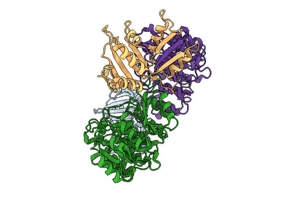 |
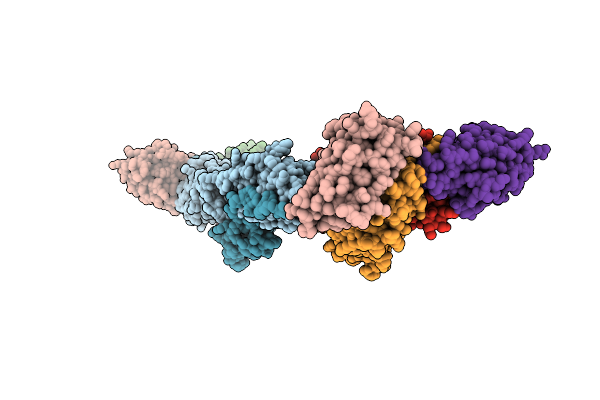
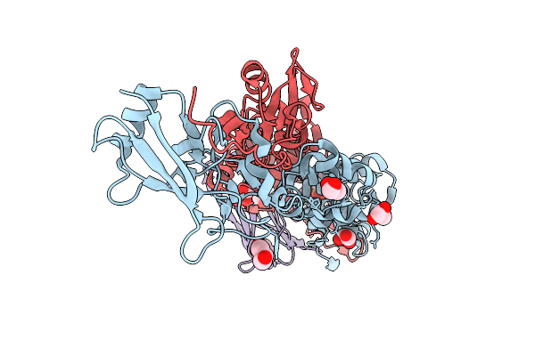
Slpl/Slph (H/L) Complex From C. Difficile Slpa (Ox247_Delta_Orf2 Strain, Slct11)
Organism: Clostridioides difficile
Method: X-RAY DIFFRACTION Release Date: 2025-09-03 Classification: STRUCTURAL PROTEIN |
 |
Organism: Clostridioides difficile
Method: X-RAY DIFFRACTION Release Date: 2025-09-03 Classification: STRUCTURAL PROTEIN |
 |
Organism: Vaccinia virus western reserve
Method: ELECTRON MICROSCOPY Release Date: 2025-09-03 Classification: VIRAL PROTEIN Ligands: NAG |
 |
Organism: Vaccinia virus western reserve
Method: X-RAY DIFFRACTION Release Date: 2025-09-03 Classification: VIRAL PROTEIN Ligands: PEG |
 |
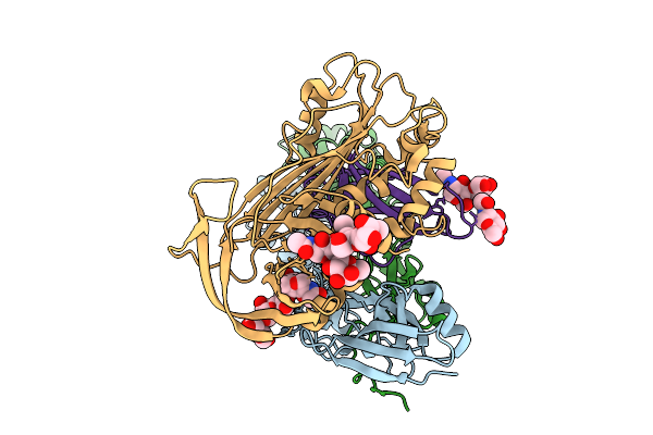
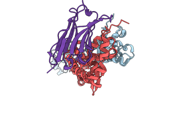
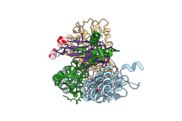
Structure Of A16/G9 (Vaccinia Virus) In Complex With Vhh D07, Vhh B01 And Vhh C05
Organism: Vaccinia virus western reserve, Vicugna pacos
Method: X-RAY DIFFRACTION Release Date: 2025-09-03 Classification: VIRAL PROTEIN Ligands: K, PO4 |
 |
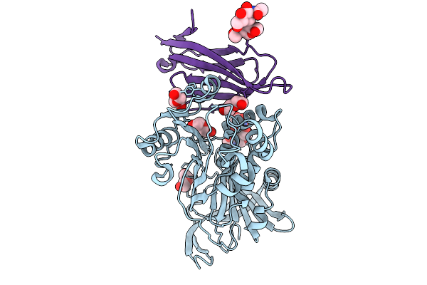
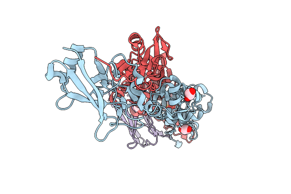
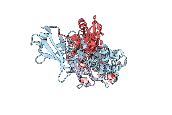
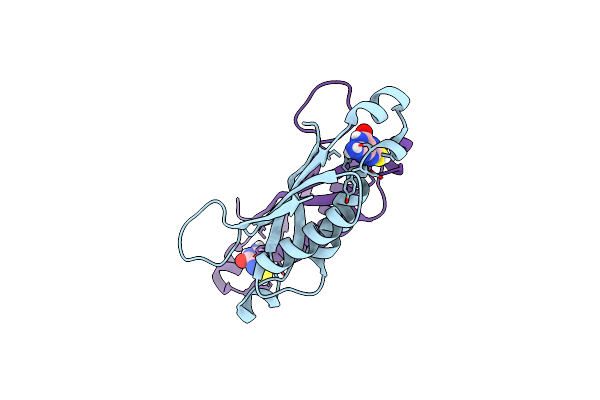
Structure Of A16/G9 (G9 Mutant - H44Y) Of Vaccinia Virus In Complex With Vhh D07
Organism: Vaccinia virus western reserve, Vicugna pacos
Method: X-RAY DIFFRACTION Release Date: 2025-09-03 Classification: VIRAL PROTEIN |
 |
Organism: Vaccinia virus western reserve, Vicugna pacos
Method: X-RAY DIFFRACTION Release Date: 2025-09-03 Classification: VIRAL PROTEIN Ligands: PO4, SO4, PEG |
 |
Organism: Vaccinia virus western reserve, Vicugna pacos
Method: X-RAY DIFFRACTION Release Date: 2025-09-03 Classification: VIRAL PROTEIN Ligands: MES, GOL |
 |
Organism: Vaccinia virus western reserve, Vicugna pacos
Method: X-RAY DIFFRACTION Release Date: 2025-09-03 Classification: VIRAL PROTEIN Ligands: MES, GOL |
 |
Organism: Vaccinia virus western reserve, Vicugna pacos
Method: X-RAY DIFFRACTION Release Date: 2025-09-03 Classification: VIRAL PROTEIN Ligands: GOL, MES |
 |
Organism: Vaccinia virus western reserve
Method: ELECTRON MICROSCOPY Release Date: 2025-09-03 Classification: VIRAL PROTEIN Ligands: NAG |
 |
Organism: Citrus sinensis, Arabidopsis thaliana
Method: X-RAY DIFFRACTION Release Date: 2025-08-20 Classification: PLANT PROTEIN Ligands: A1IRJ, MN, CL |
 |
Organism: Citrus sinensis, Arabidopsis thaliana
Method: X-RAY DIFFRACTION Release Date: 2025-08-20 Classification: PLANT PROTEIN Ligands: A1IRN, MN, CL |
 |
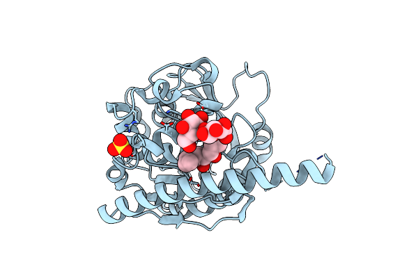
X-Ray Crystal Structure Of The Cspyl1 5M (V112L, T135L, F137I, T153I, V168A)-Icb-Hab1 Ternary Complex
Organism: Citrus sinensis, Arabidopsis thaliana
Method: X-RAY DIFFRACTION Release Date: 2025-08-20 Classification: PLANT PROTEIN Ligands: A1IRN, MN, CL |
 |
Organism: Homo sapiens
Method: X-RAY DIFFRACTION Release Date: 2025-03-12 Classification: HYDROLASE Ligands: A1H5M, EDO, PEG |
 |
Organism: Homo sapiens
Method: X-RAY DIFFRACTION Release Date: 2025-03-12 Classification: HYDROLASE Ligands: EDO, A1H5J, PEG |
 |
Organism: Mycobacterium tuberculosis
Method: X-RAY DIFFRACTION Resolution:1.95 Å Release Date: 2025-02-19 Classification: LYASE Ligands: B55 |
 |
Organism: Corynebacterium glutamicum
Method: X-RAY DIFFRACTION Resolution:2.69 Å Release Date: 2025-02-12 Classification: TRANSFERASE Ligands: VBL, SO4, CL |
 |
The Dna-Binding Domain Of L-Lactate Utilization Repressor (Lutr-Dbd) From Bacillus Subtilis
Organism: Bacillus subtilis subsp. subtilis str. 168
Method: X-RAY DIFFRACTION Resolution:1.46 Å Release Date: 2025-01-29 Classification: DNA BINDING PROTEIN |