
Search Count: 22
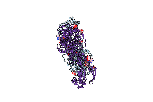 |
Organism: Pseudomonas aeruginosa (strain atcc 15692 / dsm 22644 / cip 104116 / jcm 14847 / lmg 12228 / 1c / prs 101 / pao1)
Method: X-RAY DIFFRACTION Resolution:1.73 Å Release Date: 2021-08-04 Classification: PROTEIN BINDING Ligands: VL5, CL, GOL |
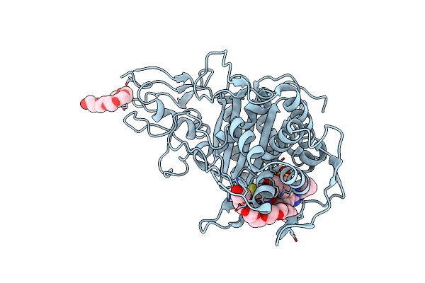 |
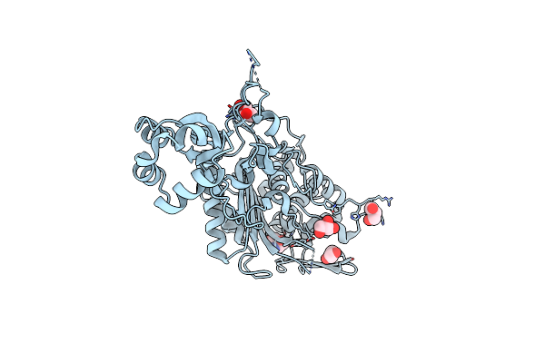
Crystal Structure Of Pbp3 Transpeptidase Domain From E. Coli In Complex With Aic499
Organism: Escherichia coli (strain k12)
Method: X-RAY DIFFRACTION Resolution:1.92 Å Release Date: 2021-08-04 Classification: PROTEIN BINDING Ligands: VL5, 12P |
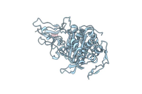 |
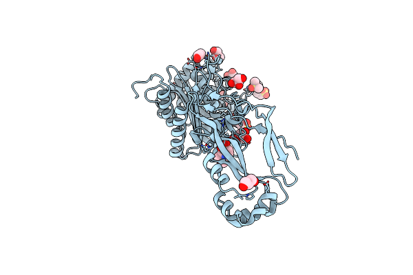
Organism: Escherichia coli (strain k12)
Method: X-RAY DIFFRACTION Resolution:2.30 Å Release Date: 2021-08-04 Classification: MEMBRANE PROTEIN Ligands: TMO |
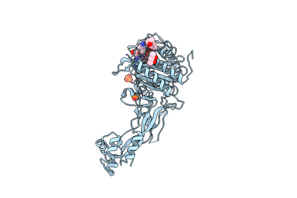 |
Organism: Escherichia coli (strain k12)
Method: X-RAY DIFFRACTION Resolution:2.70 Å Release Date: 2021-08-04 Classification: PROTEIN BINDING Ligands: VL5, PO4, MPD |
 |
Organism: Pseudomonas aeruginosa (strain atcc 15692 / dsm 22644 / cip 104116 / jcm 14847 / lmg 12228 / 1c / prs 101 / pao1)
Method: X-RAY DIFFRACTION Resolution:2.16 Å Release Date: 2021-08-04 Classification: MEMBRANE PROTEIN Ligands: GOL, TRS |
 |
Organism: Pseudomonas aeruginosa (strain atcc 15692 / dsm 22644 / cip 104116 / jcm 14847 / lmg 12228 / 1c / prs 101 / pao1)
Method: X-RAY DIFFRACTION Resolution:1.77 Å Release Date: 2021-08-04 Classification: MEMBRANE PROTEIN Ligands: GOL, MES, DMS |
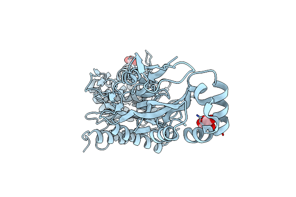 |
Organism: Pseudomonas aeruginosa (strain atcc 15692 / dsm 22644 / cip 104116 / jcm 14847 / lmg 12228 / 1c / prs 101 / pao1)
Method: X-RAY DIFFRACTION Resolution:1.86 Å Release Date: 2021-08-04 Classification: MEMBRANE PROTEIN Ligands: GOL |
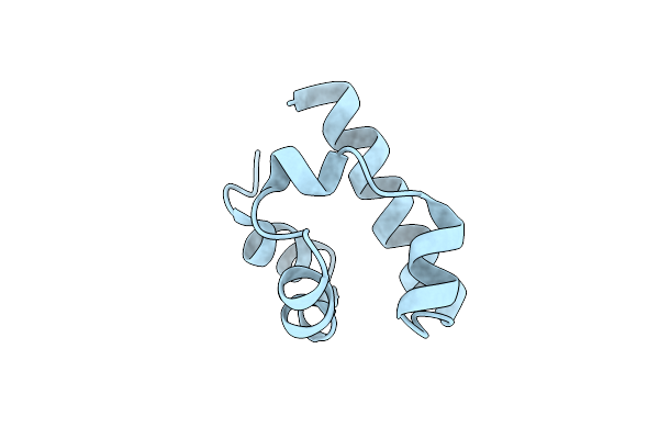 |
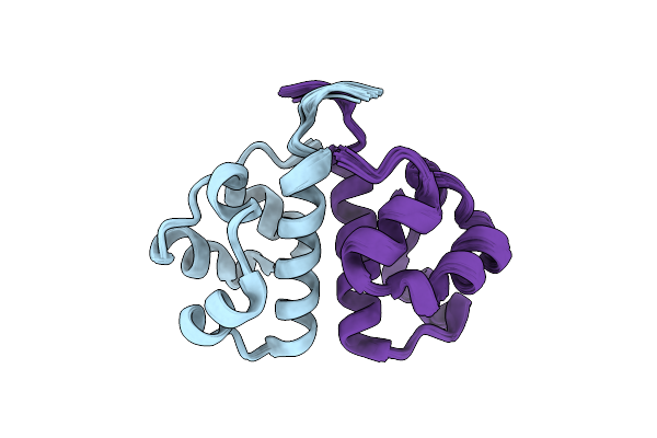
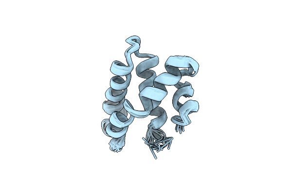
Organism: Mus musculus
Method: X-RAY DIFFRACTION Resolution:2.05 Å Release Date: 2019-01-30 Classification: SIGNALING PROTEIN |
 |
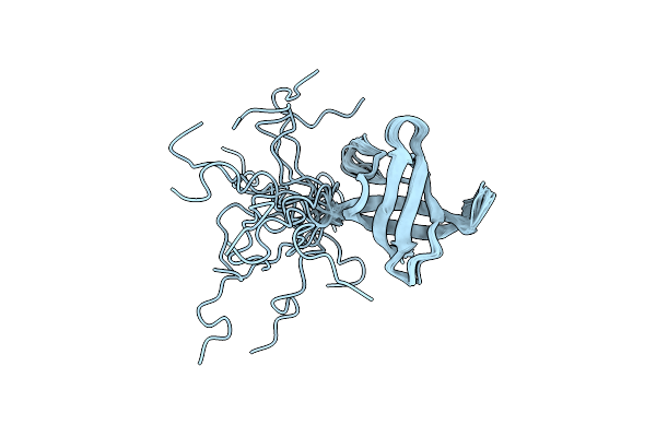
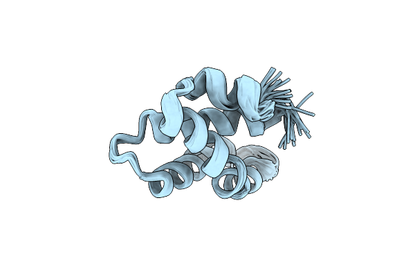
Organism: Mus musculus
Method: SOLUTION NMR Release Date: 2019-01-30 Classification: SIGNALING PROTEIN |
 |
Nmr Structure Of Cold Shock Protein A From Corynebacterium Pseudotuberculosis
Organism: Corynebacterium pseudotuberculosis
Method: SOLUTION NMR Release Date: 2017-07-19 Classification: DNA BINDING PROTEIN |
 |
 |
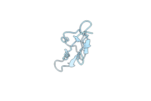
Organism: Homo sapiens
Method: SOLUTION NMR Release Date: 2013-08-28 Classification: PEPTIDE BINDING PROTEIN/PROTEIN BINDING |
 |
Solution Structure Of Venturia Inaequalis Cellophane-Induced 1 Protein (Vicin1) Domains 1 And 2
Organism: Venturia inaequalis
Method: SOLUTION NMR Release Date: 2012-07-18 Classification: CELL ADHESION |
 |
Organism: Acanthamoeba culbertsoni
Method: SOLUTION NMR Release Date: 2012-05-02 Classification: ANTIMICROBIAL PROTEIN |
 |
Organism: Acanthamoeba culbertsoni
Method: SOLUTION NMR Release Date: 2012-05-02 Classification: ANTIMICROBIAL PROTEIN |
 |
Hydramacin-1: Structure And Antibacterial Activity Of A Peptide From The Basal Metazoan Hydra
|
 |
Organism: Caenorhabditis elegans
Method: SOLUTION NMR Release Date: 2008-10-14 Classification: ANTIMICROBIAL PROTEIN |
 |
Organism: Caenorhabditis elegans
Method: SOLUTION NMR Release Date: 2008-10-14 Classification: ANTIMICROBIAL PROTEIN |
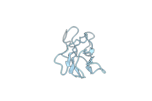 |
The Solution Structure Of The Membrane Proximal Cytokine Receptor Domain Of The Human Interleukin-6 Receptor
|
 |
Organism: Homo sapiens
Method: SOLUTION NMR Release Date: 2006-02-07 Classification: SIGNALING PROTEIN |