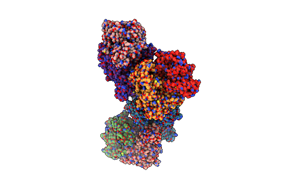
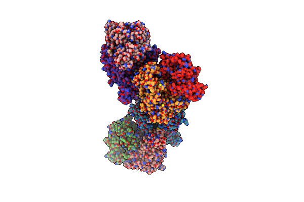
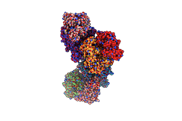
Search Count: 10
 |
Organism: Trypanosoma brucei rhodesiense
Method: X-RAY DIFFRACTION Resolution:1.60 Å Release Date: 2018-04-11 Classification: HYDROLASE Ligands: C2K, EDO |
 |
Organism: Trypanosoma brucei rhodesiense
Method: X-RAY DIFFRACTION Resolution:1.90 Å Release Date: 2018-04-11 Classification: HYDROLASE Ligands: EDO, C3E |
 |
Organism: Trypanosoma brucei rhodesiense
Method: X-RAY DIFFRACTION Resolution:2.50 Å Release Date: 2018-04-11 Classification: HYDROLASE Ligands: EDO, C2W |
 |
Cathepsin L In Complex With (3S,14E)-19-Chloro-N-(1-Cyanocyclopropyl)-5-Oxo-12,17-Dioxa-4-Azatricyclo[16.2.2.06,11]Docosa-1(21),6(11),7,9,14,18(22),19-Heptaene-3-Carboxamide
Organism: Homo sapiens
Method: X-RAY DIFFRACTION Resolution:1.37 Å Release Date: 2018-04-11 Classification: HYDROLASE Ligands: C3E, GOL |
 |
Cathepsin L In Complex With (3S,14E)-19-Chloro-N-(1-Cyanocyclopropyl)-5-Oxo-17-Oxa-4-Azatricyclo[16.2.2.06,11]Docosa-1(21),6,8,10,14,18(22),19-Heptaene-3-Carboxamide
Organism: Homo sapiens
Method: X-RAY DIFFRACTION Resolution:2.34 Å Release Date: 2018-04-11 Classification: HYDROLASE Ligands: C7Q |
 |
Cathepsin L In Complex With (3S,14E)-8-(Azetidin-3-Yl)-19-Chloro-N-(1-Cyanocyclopropyl)-5-Oxo-12,17-Dioxa-4-Azatricyclo[16.2.2.06,11]Docosa-1(21),6,8,10,14,18(22),19-Heptaene-3-Carboxamide
Organism: Homo sapiens
Method: X-RAY DIFFRACTION Resolution:2.02 Å Release Date: 2018-04-11 Classification: HYDROLASE Ligands: C7T, ZN, CL |
 |
Crystal Structure Of Xanthine Dehydrogenase (Desulfo Form) From Rhodobacter Capsulatus In Complex With Hypoxanthine
Organism: Rhodobacter capsulatus
Method: X-RAY DIFFRACTION Resolution:2.90 Å Release Date: 2008-12-23 Classification: OXIDOREDUCTASE Ligands: FES, FAD, MTE, CA, HPA, MOM |
 |
Crystal Structure Of Xanthine Dehydrogenase (Desulfo Form) From Rhodobacter Capsulatus In Complex With Xanthine
Organism: Rhodobacter capsulatus
Method: X-RAY DIFFRACTION Resolution:2.60 Å Release Date: 2008-12-23 Classification: OXIDOREDUCTASE Ligands: FES, FAD, MTE, CA, XAN, MOM |
 |
Crystal Structure Of Xanthine Dehydrogenase From Rhodobacter Capsulatus In Complex With Bound Inhibitor Pterin-6-Aldehyde
Organism: Rhodobacter capsulatus
Method: X-RAY DIFFRACTION Resolution:3.30 Å Release Date: 2008-12-23 Classification: OXIDOREDUCTASE Ligands: FES, FAD, XAX, BA, HHR |
 |
Crystal Structure Of Xanthine Dehydrogenase (E232Q Variant) From Rhodobacter Capsulatus In Complex With Hypoxanthine
Organism: Rhodobacter capsulatus
Method: X-RAY DIFFRACTION Resolution:3.40 Å Release Date: 2008-12-23 Classification: OXIDOREDUCTASE Ligands: FES, FAD, XAX, BA, HPA |

