
Search Count: 25
 |
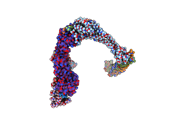
Organism: Mycolicibacterium smegmatis mc2 155
Method: ELECTRON MICROSCOPY Release Date: 2024-10-23 Classification: MEMBRANE PROTEIN Ligands: CU, HEA, CDL, HEM, MQ9, 9Y0, PLM, 9XX, HEC, 9YF, FES |
 |
Organism: Saccharomyces cerevisiae
Method: ELECTRON MICROSCOPY Release Date: 2024-10-02 Classification: MEMBRANE PROTEIN Ligands: CDL, HEM, PEF, PCF, UQ6, HEC, FES, CU, HEA, CA, MG, CUA, ZN |
 |
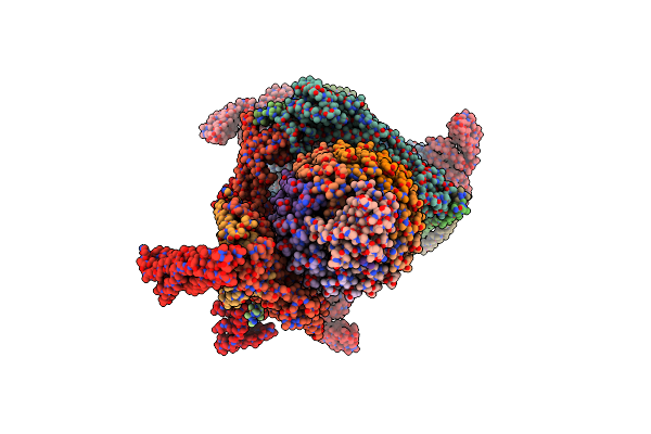
Organism: Legionella pneumophila subsp. pneumophila str. philadelphia 1, Rattus norvegicus
Method: ELECTRON MICROSCOPY Release Date: 2024-07-03 Classification: PROTON TRANSPORT Ligands: ADP |
 |
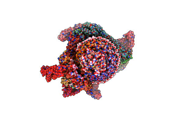
Organism: Rattus norvegicus
Method: ELECTRON MICROSCOPY Release Date: 2024-06-26 Classification: PROTON TRANSPORT Ligands: PC1, WJP, NAG, LP3, PTY, CLR |
 |
Synaptic Vesicle V-Atpase With Synaptophysin And Sidk, State 3, Peripheral Stalks
Organism: Rattus norvegicus
Method: ELECTRON MICROSCOPY Release Date: 2024-06-26 Classification: PROTON TRANSPORT |
 |
Organism: Legionella pneumophila subsp. pneumophila, Rattus norvegicus
Method: ELECTRON MICROSCOPY Release Date: 2024-06-26 Classification: PROTON TRANSPORT |
 |
Organism: Legionella pneumophila subsp. pneumophila, Rattus norvegicus
Method: ELECTRON MICROSCOPY Release Date: 2024-06-26 Classification: PROTON TRANSPORT |
 |
Organism: Legionella pneumophila subsp. pneumophila str. philadelphia 1, Rattus norvegicus
Method: ELECTRON MICROSCOPY Release Date: 2024-06-26 Classification: PROTON TRANSPORT Ligands: ADP, PC1, PTY, WJP, NAG, LP3, CLR |
 |
Organism: Pseudomonas aeruginosa
Method: ELECTRON MICROSCOPY Release Date: 2023-10-11 Classification: MEMBRANE PROTEIN Ligands: FES, HEM, U10, I7Y, HEC, CU, CA |
 |
Organism: Pseudomonas aeruginosa
Method: ELECTRON MICROSCOPY Release Date: 2023-10-11 Classification: MEMBRANE PROTEIN Ligands: CU, CA, HEM, FES, I7Y, U10, HEC |
 |
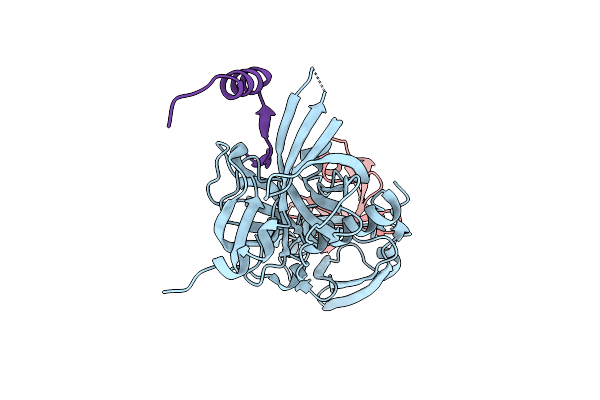
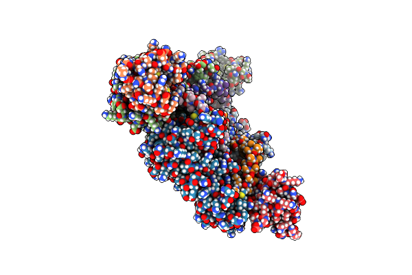
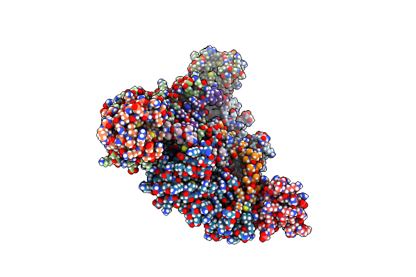
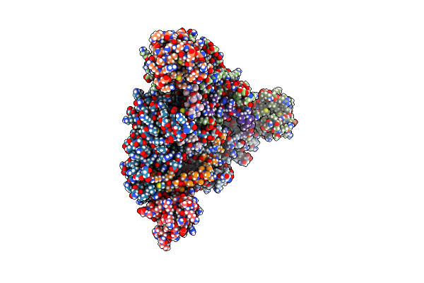
Organism: Escherichia coli (strain k12), Homo sapiens
Method: ELECTRON MICROSCOPY Release Date: 2022-11-23 Classification: CHAPERONE, HYDROLASE |
 |
Organism: Escherichia coli (strain k12), Homo sapiens
Method: ELECTRON MICROSCOPY Release Date: 2022-11-23 Classification: CHAPERONE, HYDROLASE |
 |
Organism: Escherichia coli (strain k12), Homo sapiens
Method: ELECTRON MICROSCOPY Release Date: 2022-11-23 Classification: CHAPERONE, HYDROLASE |
 |
Organism: Escherichia coli (strain k12), Homo sapiens
Method: ELECTRON MICROSCOPY Release Date: 2022-11-23 Classification: CHAPERONE, HYDROLASE |
 |
Organism: Escherichia coli (strain k12), Homo sapiens
Method: ELECTRON MICROSCOPY Release Date: 2022-11-23 Classification: CHAPERONE, HYDROLASE |
 |
Organism: Escherichia coli (strain k12), Homo sapiens
Method: ELECTRON MICROSCOPY Release Date: 2022-11-23 Classification: CHAPERONE, HYDROLASE |
 |
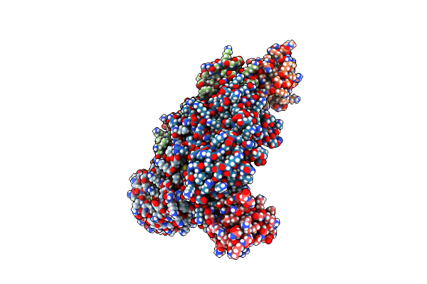
Organism: Bos taurus
Method: ELECTRON MICROSCOPY Resolution:3.10 Å Release Date: 2022-07-06 Classification: MEMBRANE PROTEIN Ligands: CU, MG, NA, HEA, PGV, PEK, 9Z9, ZN |
 |
Organism: Candida albicans (strain sc5314 / atcc mya-2876)
Method: ELECTRON MICROSCOPY Release Date: 2021-09-15 Classification: MEMBRANE PROTEIN Ligands: HEM, U10, HEC, FES |
 |
Complex Iii2 From Candida Albicans, Inhibitor Free, Rieske Head Domain In B Position
Organism: Candida albicans (strain sc5314 / atcc mya-2876)
Method: ELECTRON MICROSCOPY Release Date: 2021-09-15 Classification: MEMBRANE PROTEIN Ligands: HEM, U10, HEC, FES |
 |
Complex Iii2 From Candida Albicans, Inhibitor Free, Rieske Head Domain In Intermediate Position
Organism: Candida albicans (strain sc5314 / atcc mya-2876)
Method: ELECTRON MICROSCOPY Release Date: 2021-09-15 Classification: MEMBRANE PROTEIN Ligands: HEM, U10, HEC, FES |