
Search Count: 8
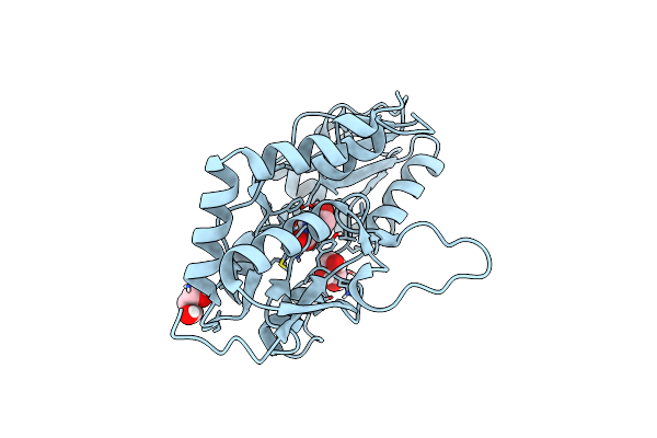 |
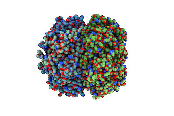
Crystal Structure Of The Drosophila Glur1A Ligand Binding Domain Y792T Mutant Complex With Glutamate
Organism: Drosophila melanogaster
Method: X-RAY DIFFRACTION Resolution:1.68 Å Release Date: 2016-12-21 Classification: MEMBRANE PROTEIN Ligands: GLU, GOL |
 |
Crystal Structure Of The Drosophila Cg3822 Kair1D Ligand Binding Domain Complex With Nmda
Organism: Drosophila melanogaster
Method: X-RAY DIFFRACTION Resolution:1.28 Å Release Date: 2016-11-09 Classification: MEMBRANE PROTEIN Ligands: OEM |
 |
Crystal Structure Of The Drosophila Cg3822 Kair1D Ligand Binding Domain Complex With D-Ap5
Organism: Drosophila melanogaster, Drosophila grimshawi
Method: X-RAY DIFFRACTION Resolution:1.75 Å Release Date: 2016-11-09 Classification: MEMBRANE PROTEIN Ligands: 2JJ, 5OY |
 |
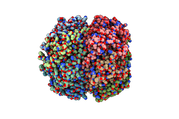
Crystal Structure Of The Drosophila Glur1A Ligand Binding Domain Complex With Glutamate
Organism: Drosophila melanogaster
Method: X-RAY DIFFRACTION Resolution:1.60 Å Release Date: 2016-09-28 Classification: MEMBRANE PROTEIN Ligands: GLU, GOL |
 |
Crystal Structure Of The Drosophila Cg3822 Kair1D Ligand Binding Domain Complex With Glutamate
Organism: Drosophila melanogaster
Method: X-RAY DIFFRACTION Resolution:1.84 Å Release Date: 2016-09-28 Classification: MEMBRANE PROTEIN Ligands: GLU, GOL, NA |
 |
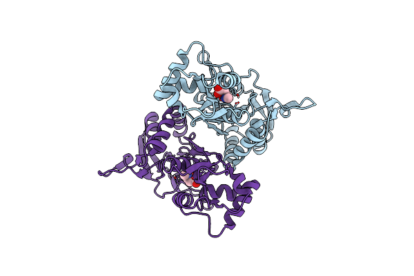
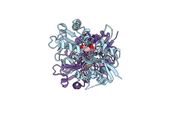
Crystal Structure Of The Active Form Of The Proteolytic Complex Clpp1 And Clpp2
Organism: Mycobacterium tuberculosis (strain cdc 1551 / oshkosh), Synthetic construct
Method: X-RAY DIFFRACTION Resolution:3.07 Å Release Date: 2016-02-17 Classification: HYDROLASE |
 |
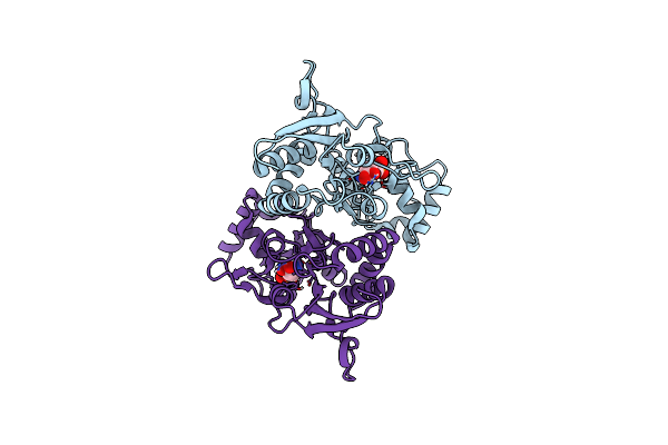
Crystal Structure Of The Active Form Of The Proteolytic Complex Clpp1 And Clpp2
Organism: Mycobacterium tuberculosis (strain cdc 1551 / oshkosh)
Method: X-RAY DIFFRACTION Resolution:2.90 Å Release Date: 2016-02-17 Classification: HYDROLASE |
 |
Organism: Drosophila melanogaster
Method: X-RAY DIFFRACTION Resolution:2.00 Å Release Date: 2015-04-29 Classification: MEMBRANE PROTEIN Ligands: GLU |