
Search Count: 109
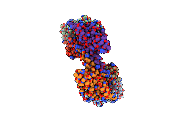 |
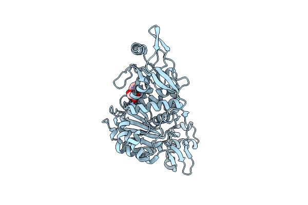
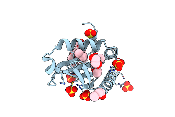
Human Antibody (Fab) And P. Aeruginosa (T3Ss) Protein Pcrv-Fragment Complex
Organism: Homo sapiens, Pseudomonas aeruginosa
Method: X-RAY DIFFRACTION Release Date: 2025-06-18 Classification: IMMUNE SYSTEM Ligands: CL, PEG |
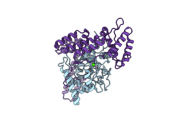 |
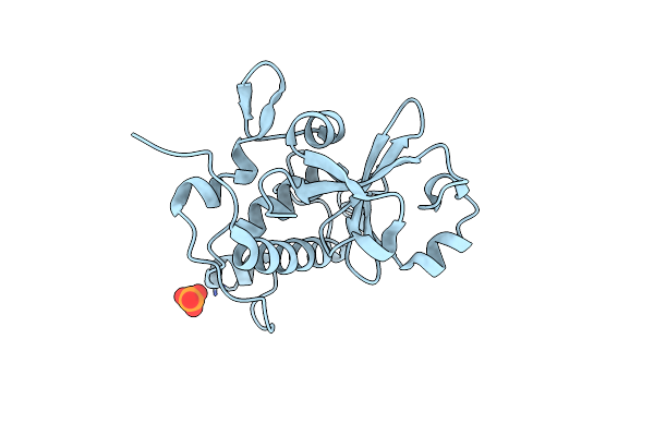
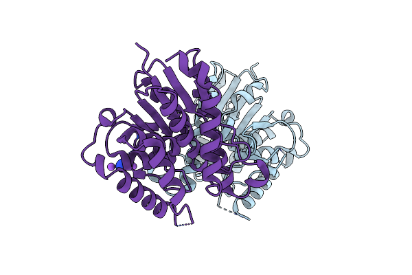
Organism: Acinetobacter baumannii
Method: X-RAY DIFFRACTION Resolution:3.17 Å Release Date: 2025-02-19 Classification: LIPID BINDING PROTEIN Ligands: CA, CL |
 |
Architecture Of The Mure-Murf Ligase Bacterial Cell Wall Biosynthesis Complex
Organism: Bordetella pertussis 18323
Method: X-RAY DIFFRACTION Resolution:2.56 Å Release Date: 2023-06-07 Classification: LIGASE Ligands: SO4 |
 |
Architecture Of A Pks-Nrps Hybrid Megaenzyme Involved In The Biosynthesis Of The Genotoxin Colibactin
Organism: Escherichia coli
Method: X-RAY DIFFRACTION Resolution:2.98 Å Release Date: 2023-04-26 Classification: BIOSYNTHETIC PROTEIN |
 |
Organism: Streptococcus pneumoniae r6
Method: X-RAY DIFFRACTION Resolution:1.47 Å Release Date: 2022-11-02 Classification: TRANSFERASE Ligands: MG, CL |
 |
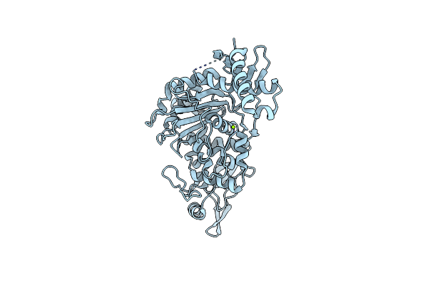
Penicillin-Binding Protein 1B (Pbp-1B) In Complex With Lactone 5Az - Streptococcus Pneumoniae R6
Organism: Streptococcus pneumoniae r6
Method: X-RAY DIFFRACTION Resolution:1.57 Å Release Date: 2022-11-02 Classification: TRANSFERASE Ligands: K2O, CL, DMS |
 |
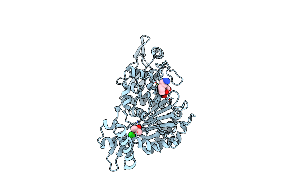
Penicillin-Binding Protein 1B (Pbp-1B) In Complex With Lactone 6Az - Streptococcus Pneumoniae R6
Organism: Streptococcus pneumoniae r6
Method: X-RAY DIFFRACTION Resolution:1.55 Å Release Date: 2022-11-02 Classification: TRANSFERASE Ligands: KQN, CL, DMS |
 |
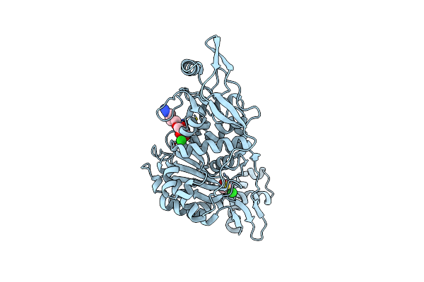
Penicillin-Binding Protein 1B (Pbp-1B) In Complex With Lactone 7Az - Streptococcus Pneumoniae R6
Organism: Streptococcus pneumoniae r6
Method: X-RAY DIFFRACTION Resolution:1.63 Å Release Date: 2022-11-02 Classification: TRANSFERASE Ligands: KQI, CL, DMS |
 |
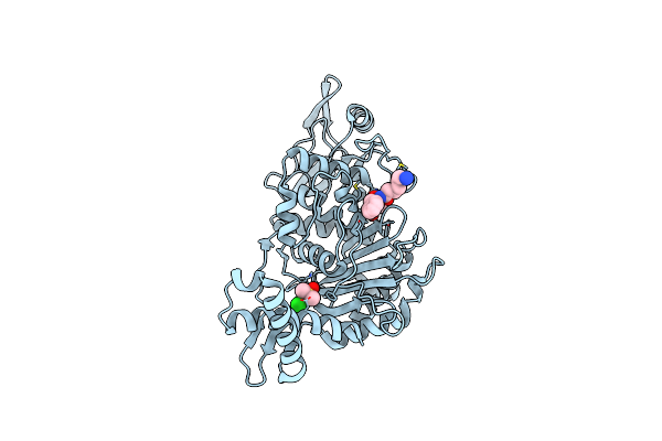
Penicillin-Binding Protein 1B (Pbp-1B) In Complex With 8Az Lactone - Streptococcus Pneumoniae R6
Organism: Streptococcus pneumoniae r6
Method: X-RAY DIFFRACTION Resolution:1.74 Å Release Date: 2022-11-02 Classification: TRANSFERASE Ligands: JWL, CL |
 |
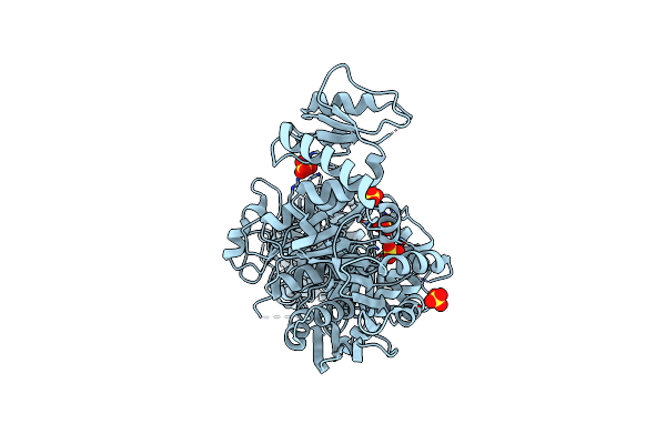
Organism: Pseudomonas aeruginosa (strain atcc 15692 / dsm 22644 / cip 104116 / jcm 14847 / lmg 12228 / 1c / prs 101 / pao1)
Method: X-RAY DIFFRACTION Resolution:1.74 Å Release Date: 2021-07-21 Classification: HYDROLASE Ligands: PO4 |
 |
Organism: Pseudomonas aeruginosa
Method: ELECTRON MICROSCOPY Resolution:3.50 Å Release Date: 2021-03-17 Classification: STRUCTURAL PROTEIN |
 |
Organism: Pseudomonas aeruginosa pao1
Method: X-RAY DIFFRACTION Resolution:1.47 Å Release Date: 2021-03-17 Classification: STRUCTURAL PROTEIN Ligands: MG, CL |
 |
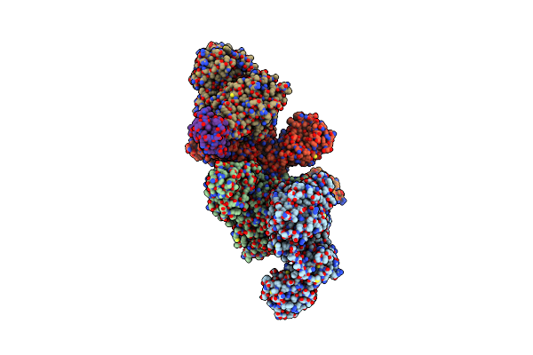
Organism: Aeromonas hydrophila
Method: ELECTRON MICROSCOPY Resolution:3.70 Å Release Date: 2019-04-10 Classification: PROTEIN TRANSPORT |
 |
Organism: Vibrio vulnificus
Method: ELECTRON MICROSCOPY Resolution:3.40 Å Release Date: 2019-04-10 Classification: PROTEIN TRANSPORT |
 |
Organism: Vibrio vulnificus
Method: X-RAY DIFFRACTION Resolution:1.75 Å Release Date: 2019-04-10 Classification: PROTEIN BINDING Ligands: 1PE, EDO, SO3, SO4 |
 |
Organism: Helicobacter pylori (strain atcc 700392 / 26695)
Method: X-RAY DIFFRACTION Resolution:3.03 Å Release Date: 2017-08-23 Classification: HYDROLASE/ANTIBIOTIC Ligands: SO4 |
 |
Complex Between Penicillin-Binding Protein (Pbp2) And Mrec From Helicobacter Pylori
Organism: Helicobacter pylori (strain atcc 700392 / 26695)
Method: X-RAY DIFFRACTION Resolution:2.74 Å Release Date: 2017-08-23 Classification: HYDROLASE/ANTIBIOTIC |
 |
Organism: Escherichia coli k-12
Method: X-RAY DIFFRACTION Resolution:1.84 Å Release Date: 2016-04-13 Classification: LIGASE Ligands: MPD, NI, SO4 |
 |
Crystal Structure Of Murd Ligase From Escherichia Coli In Complex With Uma And Adp
Organism: Escherichia coli k-12
Method: X-RAY DIFFRACTION Resolution:1.90 Å Release Date: 2016-04-13 Classification: LIGASE Ligands: UMA, ADP, MLI |
 |
Organism: Pseudomonas aeruginosa pao1
Method: X-RAY DIFFRACTION Resolution:2.74 Å Release Date: 2016-04-06 Classification: TRANSPORT PROTEIN Ligands: AZI, NA |