
Search Count: 31
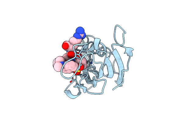 |
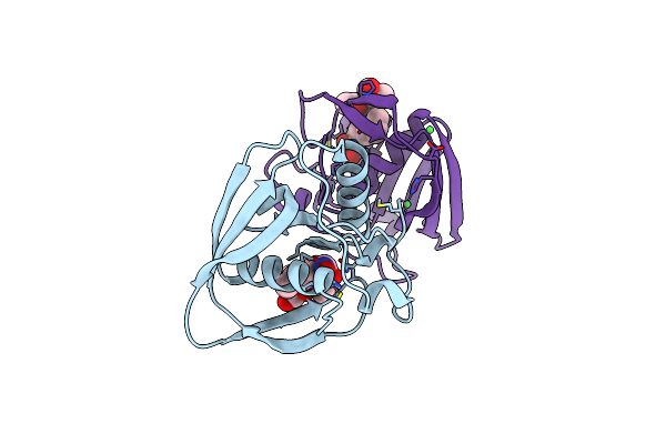
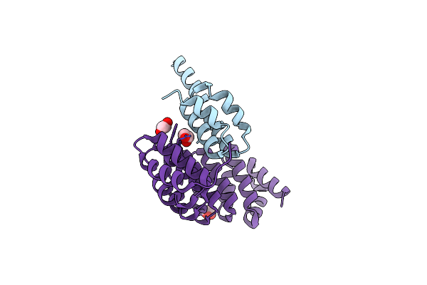
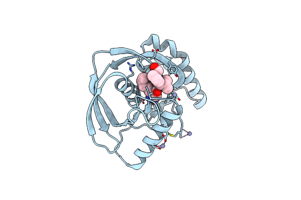
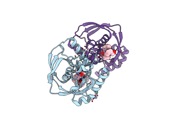
Crystal Structure Of Fk1 Domain Of Fkbp52 In Complex With A Bio-Inspired Hybrid Fluorescent Ligand
Organism: Homo sapiens
Method: X-RAY DIFFRACTION Resolution:2.30 Å Release Date: 2020-05-13 Classification: ISOMERASE Ligands: K0T |
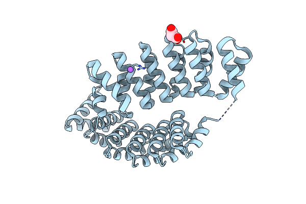 |
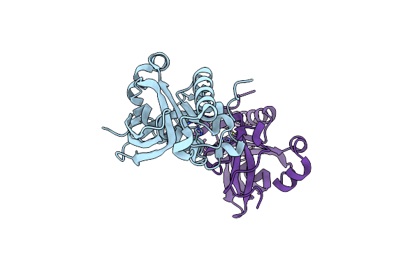
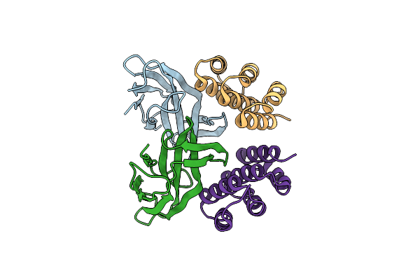
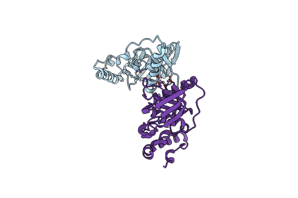
Structure Of A3_Bgfpd, An Artificial Bi-Domain Protein Based On Two Different Alpharep Domains : A3 And A Gfp Binding Domain (Bgfpd)
Organism: Synthetic construct
Method: X-RAY DIFFRACTION Resolution:2.55 Å Release Date: 2018-10-24 Classification: BIOSYNTHETIC PROTEIN Ligands: MLI, NA |
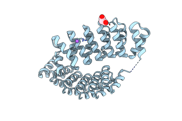 |
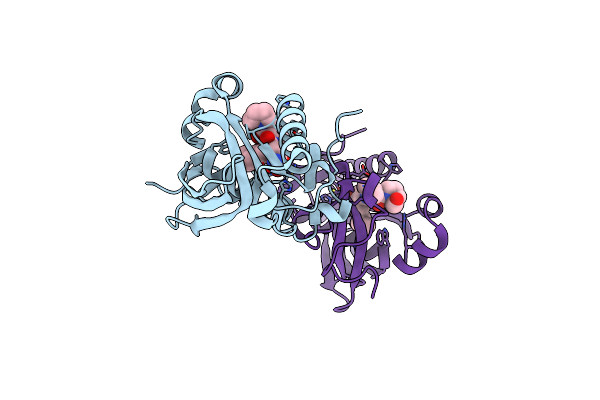
Structure Of A3_Bgfpd, An Artificial Bi-Domain Protein Based On Two Different Alpharep Domains : A3 And A Gfp Binding Domain (Bgfpd)
Organism: Synthetic construct
Method: X-RAY DIFFRACTION Resolution:2.79 Å Release Date: 2018-08-08 Classification: BIOSYNTHETIC PROTEIN Ligands: MLI, NA |
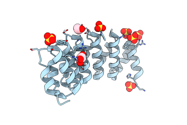 |
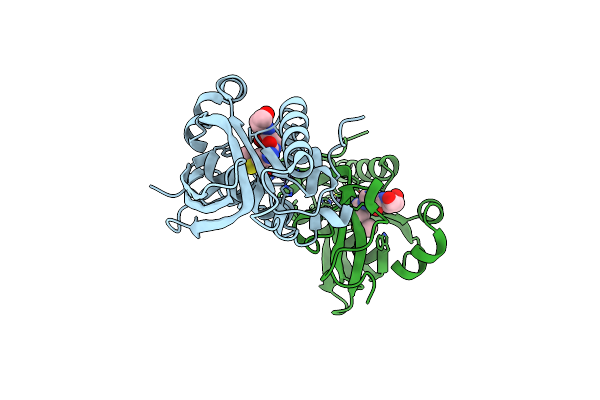
Structure Of A3_A3, An Artificial Bi-Domain Protein Based On Two Identical Alpharep A3 Domains
Organism: Synthetic construct
Method: X-RAY DIFFRACTION Resolution:1.94 Å Release Date: 2018-08-08 Classification: BIOSYNTHETIC PROTEIN Ligands: GOL, SO4 |
 |
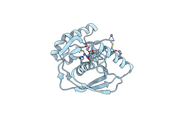
Crystal Structure Of Pdf From The Vibrio Parahaemolyticus Bacteriophage Vp16T In Complex With Actinonin - Crystal Form Ii
Organism: Vibrio phage vp16t
Method: X-RAY DIFFRACTION Resolution:1.40 Å Release Date: 2017-11-29 Classification: HYDROLASE Ligands: BB2, ZN, NI |
 |
Organism: Aequorea victoria, Synthetic construct
Method: X-RAY DIFFRACTION Resolution:2.00 Å Release Date: 2015-08-19 Classification: PROTEIN BINDING |
 |
Organism: Aequorea victoria, Synthetic construct
Method: X-RAY DIFFRACTION Resolution:3.40 Å Release Date: 2015-08-19 Classification: PROTEIN BINDING |
 |
Organism: Arabidopsis thaliana
Method: X-RAY DIFFRACTION Resolution:2.00 Å Release Date: 2014-02-26 Classification: HYDROLASE Ligands: ZN |
 |
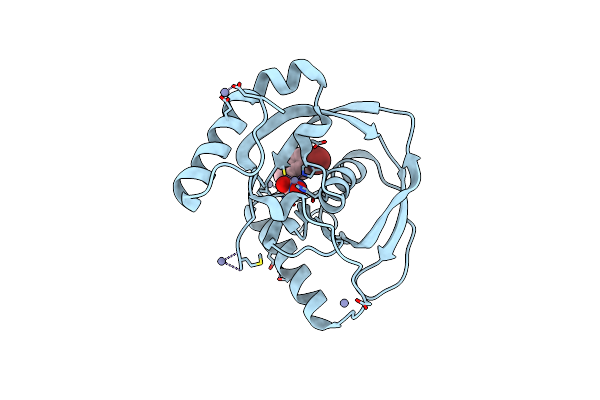
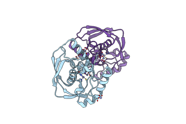
Crystal Structure Of A Human-Like Mitochondrial Peptide Deformylase In Complex With Actinonin
Organism: Arabidopsis thaliana
Method: X-RAY DIFFRACTION Resolution:2.10 Å Release Date: 2014-02-26 Classification: HYDROLASE/HYDROLASE INHIBITOR Ligands: BB2, ZN |
 |
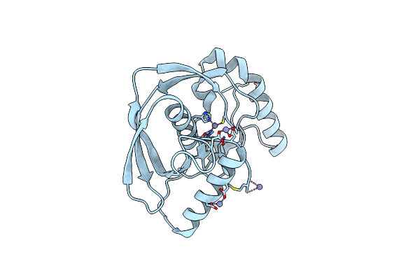
Crystal Structure Of A Human-Like Mitochondrial Peptide Deformylase In Complex With Met-Ala-Ser
Organism: Arabidopsis thaliana
Method: X-RAY DIFFRACTION Resolution:2.40 Å Release Date: 2014-02-26 Classification: HYDROLASE/PEPTIDE Ligands: ZN |
 |
Selection Of Specific Protein Binders For Pre-Defined Targets From An Optimized Library Of Artificial Helicoidal Repeat Proteins (Alpharep)
Organism: Synthetic construct
Method: X-RAY DIFFRACTION Resolution:1.90 Å Release Date: 2013-09-25 Classification: DE NOVO PROTEIN/protein binding Ligands: EDO |
 |
Selection Of Specific Protein Binders For Pre-Defined Targets From An Optimized Library Of Artificial Helicoidal Repeat Proteins (Alpharep)
Organism: Streptomyces malayensis, Synthetic construct
Method: X-RAY DIFFRACTION Resolution:2.60 Å Release Date: 2013-09-25 Classification: DNA binding protein/de novo protein |
 |
Crystal Structure Of Arabidopsis Thaliana Peptide Deformylase 1B (Atpdf1B) In Complex With Inhibitor 6B
Organism: Arabidopsis thaliana
Method: X-RAY DIFFRACTION Resolution:3.00 Å Release Date: 2011-06-08 Classification: HYDROLASE/HYDROLASE INHIBITOR Ligands: ZN, BB4 |
 |
Crystal Structure Of Arabidopsis Thaliana Petide Deformylase 1B (Atpdf1B) (Crystallized In Peg-550-Mme)
Organism: Arabidopsis thaliana
Method: X-RAY DIFFRACTION Resolution:2.00 Å Release Date: 2011-06-08 Classification: HYDROLASE Ligands: ZN |
 |
Crystal Structure Of Arabidopsis Thaliana Petide Deformylase 1B (Atpdf1B) In Complex With Inhibitor 21
Organism: Arabidopsis thaliana
Method: X-RAY DIFFRACTION Resolution:1.30 Å Release Date: 2011-06-08 Classification: Hydrolase/Hydrolase Inhibitor Ligands: PN3, ZN |
 |
Crystal Structure Of Arabidopsis Thaliana Petide Deformylase 1B (Atpdf1B) In Complex With Actinonin (Crystallized In Peg-550-Mme)
Organism: Arabidopsis thaliana
Method: X-RAY DIFFRACTION Resolution:1.90 Å Release Date: 2011-06-08 Classification: HYDROLASE/ANTIBIOTIC Ligands: BB2, ZN |
 |
Crystal Structure Of Arabidopsis Thaliana Petide Deformylase 1B (Atpdf1B) G41Q Mutant
Organism: Arabidopsis thaliana
Method: X-RAY DIFFRACTION Resolution:2.30 Å Release Date: 2011-06-08 Classification: HYDROLASE Ligands: ZN |
 |
Crystal Structure Of Arabidopsis Thaliana Petide Deformylase 1B (Atpdf1B) G41M Mutant
Organism: Arabidopsis thaliana
Method: X-RAY DIFFRACTION Resolution:2.10 Å Release Date: 2011-06-08 Classification: HYDROLASE Ligands: ZN |
 |
Organism: Arabidopsis thaliana
Method: X-RAY DIFFRACTION Resolution:2.00 Å Release Date: 2011-03-30 Classification: HYDROLASE Ligands: ZN |
 |
Crystal Structure Of Arabidopsis Thaliana Peptide Deformylase 1B (Atpdf1B) In Complex With Actinonin
Organism: Arabidopsis thaliana
Method: X-RAY DIFFRACTION Resolution:2.00 Å Release Date: 2011-03-30 Classification: HYDROLASE/ANTIBIOTIC Ligands: BB2, ZN |