
Search Count: 22
 |
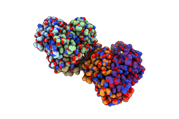
Organism: Mammalian orthoreovirus 1
Method: X-RAY DIFFRACTION Resolution:3.00 Å Release Date: 2024-03-06 Classification: RNA BINDING PROTEIN |
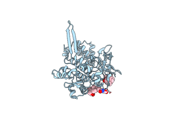 |
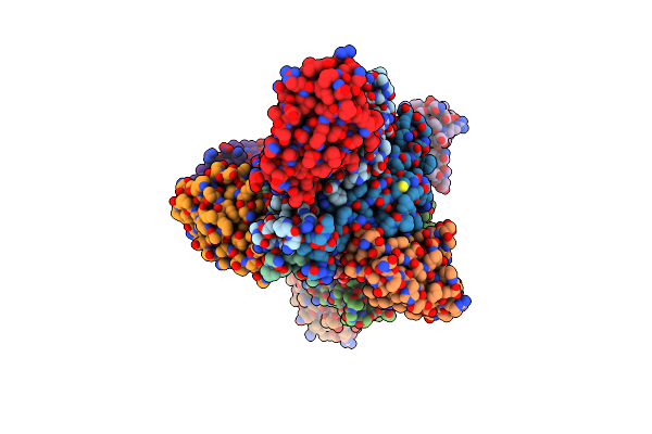
Organism: Orthoreovirus
Method: X-RAY DIFFRACTION Resolution:3.16 Å Release Date: 2024-03-06 Classification: RNA BINDING PROTEIN Ligands: GCH |
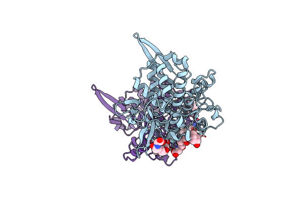 |
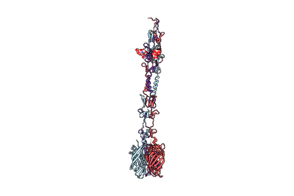
Structure Of Orthoreovirus Rna Chaperone Sigmans R6A Mutant In Complex With Bile Acid
Organism: Orthoreovirus
Method: X-RAY DIFFRACTION Resolution:3.20 Å Release Date: 2024-03-06 Classification: RNA BINDING PROTEIN Ligands: GCH |
 |
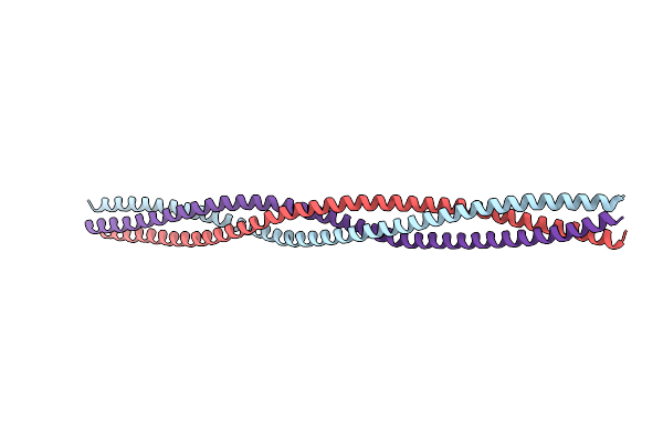
Organism: Homo sapiens
Method: ELECTRON MICROSCOPY Release Date: 2021-03-03 Classification: CHAPERONE |
 |
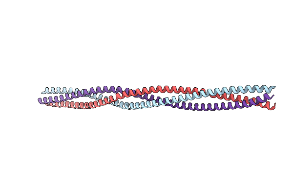
Organism: Homo sapiens, Reovirus type 3 (strain dearing)
Method: ELECTRON MICROSCOPY Release Date: 2021-03-03 Classification: CHAPERONE Ligands: ZN |
 |
Organism: Mammalian orthoreovirus 1 lang
Method: X-RAY DIFFRACTION Resolution:1.35 Å Release Date: 2018-04-25 Classification: VIRAL PROTEIN Ligands: IOD, CL |
 |
Organism: Mammalian orthoreovirus 1 lang
Method: X-RAY DIFFRACTION Resolution:1.43 Å Release Date: 2018-04-25 Classification: VIRAL PROTEIN Ligands: CL |
 |
Organism: Mammalian orthoreovirus 1 lang
Method: X-RAY DIFFRACTION Resolution:2.10 Å Release Date: 2018-04-25 Classification: VIRAL PROTEIN Ligands: CL |
 |
Organism: Mammalian orthoreovirus 3 dearing
Method: X-RAY DIFFRACTION Resolution:2.15 Å Release Date: 2018-04-25 Classification: VIRAL PROTEIN |
 |
Organism: Reovirus sp., Mus musculus
Method: X-RAY DIFFRACTION Resolution:3.00 Å Release Date: 2017-02-15 Classification: IMMUNE SYSTEM |
 |
Organism: Reovirus type 1 (strain lang), Mus musculus
Method: X-RAY DIFFRACTION Resolution:3.70 Å Release Date: 2016-12-21 Classification: IMMUNE SYSTEM |
 |
Organism: Mammalian orthoreovirus 1 lang
Method: X-RAY DIFFRACTION Resolution:2.20 Å Release Date: 2015-04-01 Classification: VIRAL PROTEIN Ligands: CL, MG, ACT, GOL |
 |
Crystal Structure Of The T1L Reovirus Attachment Protein Sigma1 In Complex With The Gm2 Glycan
Organism: Mammalian orthoreovirus 1
Method: X-RAY DIFFRACTION Resolution:3.60 Å Release Date: 2012-12-05 Classification: VIRAL PROTEIN |
 |
Crystal Structure Of The T1L Reovirus Attachment Protein Sigma1 In Complex With Alpha-2,3-Sialyllactose
Organism: Mammalian orthoreovirus 1
Method: X-RAY DIFFRACTION Resolution:3.50 Å Release Date: 2012-12-05 Classification: VIRAL PROTEIN |
 |
Structure Of Reovirus Attachment Protein Sigma1 In Complex With Alpha-2,3-Sialyllactose
Organism: Reovirus type 3
Method: X-RAY DIFFRACTION Resolution:2.25 Å Release Date: 2011-11-23 Classification: VIRAL PROTEIN |
 |
Structure Of Reovirus Attachment Protein Sigma1 In Complex With Alpha-2,6-Sialyllactose
Organism: Reovirus type 3
Method: X-RAY DIFFRACTION Resolution:2.79 Å Release Date: 2011-11-23 Classification: VIRAL PROTEIN Ligands: SIA |
 |
Structure Of Reovirus Attachment Protein Sigma1 In Complex With Alpha-2,8-Disialyllactose
Organism: Reovirus type 3
Method: X-RAY DIFFRACTION Resolution:2.28 Å Release Date: 2011-11-23 Classification: VIRAL PROTEIN |
 |
Structure Of Reovirus Sigma1 In Complex With Its Receptor Junctional Adhesion Molecule-A
Organism: Reovirus, Homo sapiens
Method: X-RAY DIFFRACTION Resolution:3.40 Å Release Date: 2008-11-04 Classification: VIRAL PROTEIN/CELL ADHESION |
 |
Crystal Structure Of Reovirus T3D Attachment Protein Sigma1 Head Domain Wild-Type At 1.75 A Resolution
Organism: Reovirus sp.
Method: X-RAY DIFFRACTION Resolution:1.75 Å Release Date: 2007-02-13 Classification: VIRAL PROTEIN Ligands: MG, GOL |
 |
Crystal Structure Of Reovirus T3D Attachment Protein Sigma1 Head Domain D345N Mutant
Organism: Reovirus sp.
Method: X-RAY DIFFRACTION Resolution:1.85 Å Release Date: 2007-02-13 Classification: VIRAL PROTEIN Ligands: MG |