
Search Count: 272
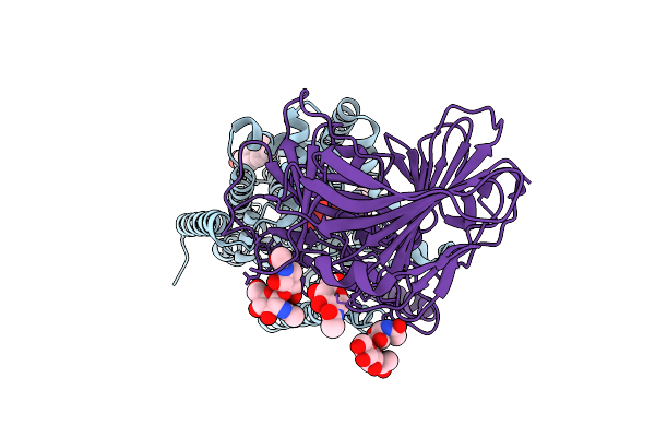 |
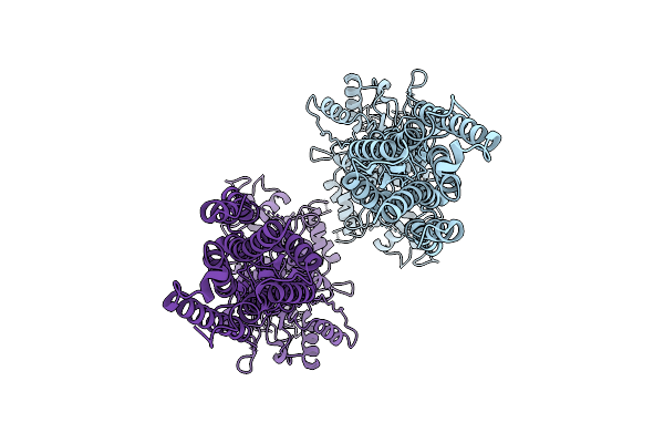
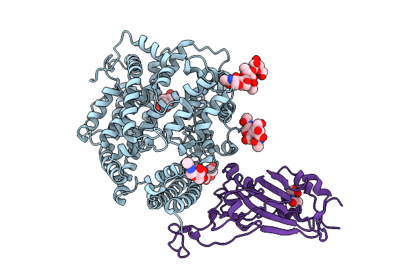
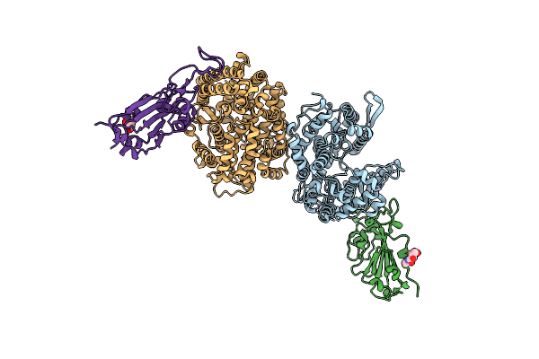
Cryo-Em Structure Of Candida Glabrata Gpi Mannosyltransferase I Bound To Dol-P-Man
Organism: Nakaseomyces glabratus
Method: ELECTRON MICROSCOPY Release Date: 2025-12-03 Classification: MEMBRANE PROTEIN Ligands: MJC, Y01, NAG |
 |
Organism: Homo sapiens
Method: ELECTRON MICROSCOPY Release Date: 2025-10-01 Classification: MEMBRANE PROTEIN Ligands: HVG, A1ENS |
 |
Organism: Homo sapiens, Mus musculus
Method: ELECTRON MICROSCOPY Release Date: 2025-10-01 Classification: MEMBRANE PROTEIN Ligands: GLU |
 |
Organism: Homo sapiens
Method: ELECTRON MICROSCOPY Release Date: 2025-10-01 Classification: MEMBRANE PROTEIN Ligands: Z99 |
 |
Organism: Homo sapiens
Method: ELECTRON MICROSCOPY Release Date: 2025-10-01 Classification: MEMBRANE PROTEIN Ligands: A1ENR, HVG |
 |
Organism: Homo sapiens
Method: ELECTRON MICROSCOPY Release Date: 2025-10-01 Classification: MEMBRANE PROTEIN |
 |
Crystal Structure Of Ratg13 Receptor-Binding Domain Complexed With Squirrel Ace2
Organism: Petaurus norfolcensis, Bat coronavirus ratg13
Method: X-RAY DIFFRACTION Release Date: 2025-08-06 Classification: VIRAL PROTEIN Ligands: CL, NAG, ZN |
 |
Crystal Structure Of Pcov-Gd Receptor-Binding Domain Complexed With Squirrel Ace2
Organism: Pangolin coronavirus, Petaurus norfolcensis
Method: X-RAY DIFFRACTION Release Date: 2025-08-06 Classification: VIRAL PROTEIN Ligands: NAG |
 |
Crystal Structure Of Pcov-Gx Receptor-Binding Domain Complexed With Squirrel Ace2
Organism: Petaurus norfolcensis, Pangolin coronavirus
Method: X-RAY DIFFRACTION Release Date: 2025-08-06 Classification: VIRAL PROTEIN Ligands: NAG |
 |
Crystal Structure Of Sars-Cov-2 Receptor-Binding Domain Complexed With Squirrel Ace2
Organism: Petaurus norfolcensis, Severe acute respiratory syndrome coronavirus 2
Method: X-RAY DIFFRACTION Release Date: 2025-08-06 Classification: VIRAL PROTEIN Ligands: NAG, ZN |
 |
Crystal Structure Of Sars-Cov-2 Jn.1 Variant Rbd Complexed With Squirrel Ace2
Organism: Petaurus norfolcensis, Severe acute respiratory syndrome coronavirus 2
Method: X-RAY DIFFRACTION Release Date: 2025-08-06 Classification: VIRAL PROTEIN/HYDROLASE Ligands: NAG, ZN |
 |
Organism: Bat coronavirus
Method: X-RAY DIFFRACTION Release Date: 2025-05-14 Classification: VIRAL PROTEIN Ligands: NAG |
 |
Organism: Xanthomonas phage phixacjx1
Method: ELECTRON MICROSCOPY Release Date: 2025-05-07 Classification: VIRUS |
 |
The Composite Cryo-Em Structure Of The Head-To-Tail Connector And Head-Proximal Tail Components Of Bacteriophage Phixacjx1
Organism: Xanthomonas phage phixacjx1
Method: ELECTRON MICROSCOPY Release Date: 2025-05-07 Classification: VIRUS |
 |
Organism: Bat coronavirus ratg13, Rhinolophus affinis
Method: X-RAY DIFFRACTION Resolution:3.01 Å Release Date: 2025-04-30 Classification: VIRAL PROTEIN/HYDROLASE Ligands: NAG |
 |
Organism: Rhinolophus affinis, Severe acute respiratory syndrome coronavirus 2
Method: X-RAY DIFFRACTION Resolution:3.60 Å Release Date: 2025-04-30 Classification: VIRAL PROTEIN/HYDROLASE Ligands: NAG |
 |
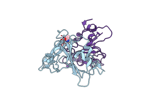
Organism: Homo sapiens, Mus musculus
Method: ELECTRON MICROSCOPY Release Date: 2025-04-23 Classification: MEMBRANE PROTEIN/IMMUNE SYSTEM Ligands: A1D5N |
 |
Organism: Streptomyces lasalocidi
Method: X-RAY DIFFRACTION Resolution:1.85 Å Release Date: 2025-04-16 Classification: FLAVOPROTEIN Ligands: FAD, GOL, CL |
 |
Organism: Streptomyces virginiae
Method: X-RAY DIFFRACTION Resolution:2.00 Å Release Date: 2025-04-16 Classification: FLAVOPROTEIN Ligands: FAD, Y7R, CL |
 |
Organism: Streptomyces virginiae
Method: X-RAY DIFFRACTION Resolution:2.10 Å Release Date: 2025-04-16 Classification: FLAVOPROTEIN Ligands: FAD, GOL, YGK, CL |