
Search Count: 48
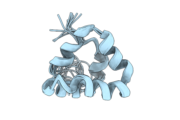 |
Organism: Homo sapiens
Method: SOLUTION NMR Release Date: 2025-10-01 Classification: METAL BINDING PROTEIN |
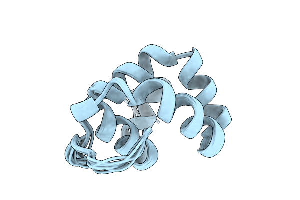 |
Organism: Homo sapiens
Method: SOLUTION NMR Release Date: 2025-10-01 Classification: METAL BINDING PROTEIN |
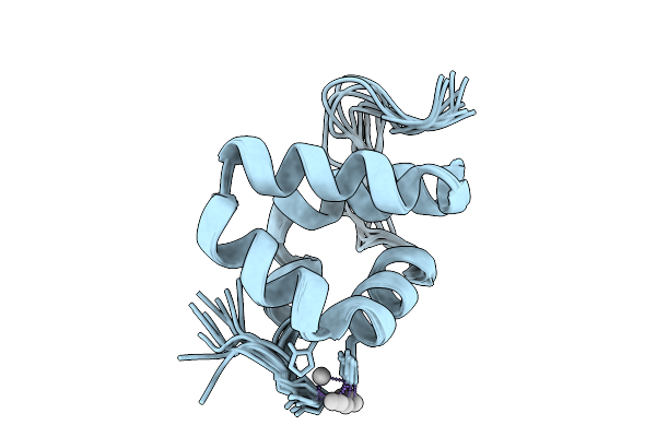 |
Solution Structure Of Silver Bound Xpc Binding Domain Of Hhr23B (Holo Form)
Organism: Homo sapiens
Method: SOLUTION NMR Release Date: 2025-10-01 Classification: METAL BINDING PROTEIN Ligands: AG |
 |
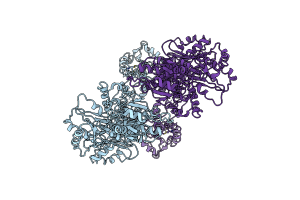
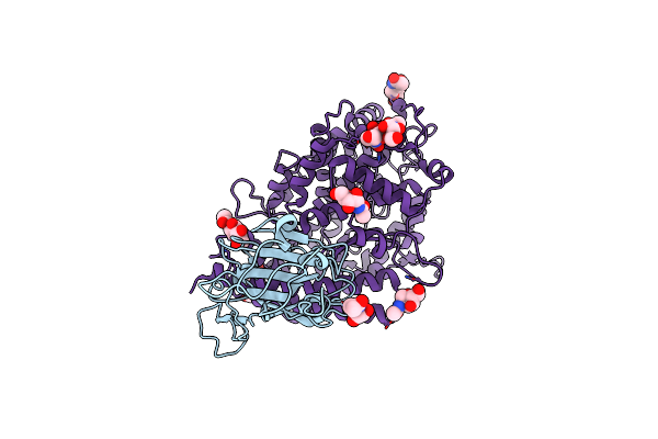
Crystal Structure Of The Lsd1/Corest Histone Demethylase In Complex With The Cofactor Fad And The Inhibitor Gsk690
Organism: Homo sapiens
Method: X-RAY DIFFRACTION Release Date: 2025-09-17 Classification: GENE REGULATION Ligands: FAD, GOL, A1A5U |
 |
Organism: Homo sapiens
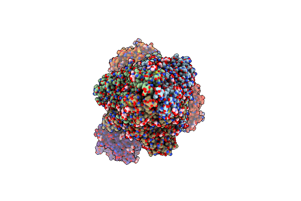
Method: ELECTRON MICROSCOPY Release Date: 2025-06-18 Classification: HYDROLASE Ligands: ZN |
 |
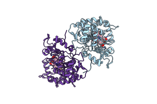
Crystal Structure Of Polyketide Synthase (Pks) Thioreductase Domain From Streptomyces Coelicolor
Organism: Streptomyces coelicolor
Method: X-RAY DIFFRACTION Release Date: 2024-11-27 Classification: BIOSYNTHETIC PROTEIN Ligands: NDP, GOL, BTB |
 |
Organism: Severe acute respiratory syndrome coronavirus 2, Homo sapiens
Method: ELECTRON MICROSCOPY Release Date: 2024-02-28 Classification: VIRAL PROTEIN/HYDROLASE Ligands: NAG |
 |
Organism: Severe acute respiratory syndrome coronavirus 2, Homo sapiens
Method: ELECTRON MICROSCOPY Release Date: 2024-02-28 Classification: VIRAL PROTEIN/HYDROLASE Ligands: NAG, ZN |
 |
Organism: Metagenome
Method: X-RAY DIFFRACTION Resolution:1.53 Å Release Date: 2023-05-10 Classification: BIOSYNTHETIC PROTEIN Ligands: HEM |
 |
Organism: Homo sapiens
Method: X-RAY DIFFRACTION Resolution:1.72 Å Release Date: 2023-01-18 Classification: TRANSPORT PROTEIN Ligands: SO4 |
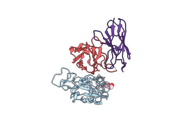 |
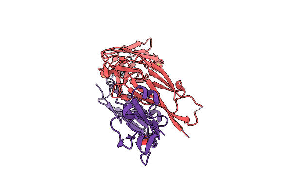
Cryo-Em Structure Of The Omicron Rbd In Complex With 35B5 Fab( Local Refinement Of The Rbd And 35B5 Fab)
Organism: Severe acute respiratory syndrome coronavirus 2, Homo sapiens
Method: ELECTRON MICROSCOPY Resolution:3.35 Å Release Date: 2022-08-17 Classification: VIRAL PROTEIN,IMMUNE SYSTEM Ligands: NAG |
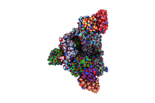 |
Cryo-Em Structure Of The Omicron S In Complex With 35B5 Fab(1 Down- And 2 Up Rbds)
Organism: Severe acute respiratory syndrome coronavirus 2, Homo sapiens
Method: ELECTRON MICROSCOPY Release Date: 2022-05-25 Classification: VIRAL PROTEIN,IMMUNE SYSTEM Ligands: NAG |
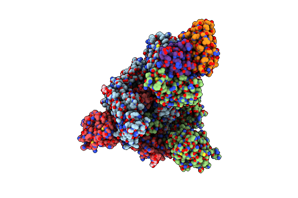 |
Cryo-Em Structure Of The Omicron S In Complex With 35B5 Fab(1 Down-, 1 Up- And 1 Invisible Rbds)
Organism: Severe acute respiratory syndrome coronavirus 2, Homo sapiens
Method: ELECTRON MICROSCOPY Release Date: 2022-05-25 Classification: VIRAL PROTEIN,IMMUNE SYSTEM Ligands: NAG |
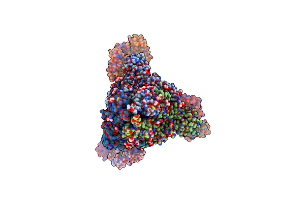 |
Cryo-Em Structure Of The Sars-Cov-2 S-6P In Complex With 35B5 Fab(1 Down Rbd, State1)
Organism: Severe acute respiratory syndrome coronavirus 2, Homo sapiens
Method: ELECTRON MICROSCOPY Release Date: 2022-04-06 Classification: VIRAL PROTEIN,IMMUNE SYSTEM Ligands: NAG |
 |
Organism: Severe acute respiratory syndrome coronavirus 2, Homo sapiens
Method: ELECTRON MICROSCOPY Release Date: 2022-04-06 Classification: VIRAL PROTEIN Ligands: NAG |
 |
Cryo-Em Structure Of The Sars-Cov-2 S-6P In Complex With Fab30 (Local Refinement Of The Rbd And Fab30)
Organism: Homo sapiens, Severe acute respiratory syndrome coronavirus 2
Method: ELECTRON MICROSCOPY Release Date: 2022-04-06 Classification: VIRAL PROTEIN |
 |
Cryo-Em Structure Of The Sars-Cov-2 S-6P In Complex With 35B5 Fab(State2, Local Refinement Of The Rbd And 35B5 Fab)
Organism: Severe acute respiratory syndrome coronavirus 2, Homo sapiens
Method: ELECTRON MICROSCOPY Release Date: 2022-03-23 Classification: VIRAL PROTEIN, IMMUNE SYSTEM Ligands: NAG |
 |
Cryo-Em Structure Of The Sars-Cov-2 S-6P In Complex With 35B5 Fab (State1, Local Refinement Of The Rbd, Ntd And 35B5 Fab)
Organism: Severe acute respiratory syndrome coronavirus 2, Homo sapiens
Method: ELECTRON MICROSCOPY Release Date: 2022-03-23 Classification: VIRAL PROTEIN Ligands: NAG |
 |
Cryo-Em Structure Of The Sars-Cov-2 S-6P In Complex With 35B5 Fab(3 Up Rbds, State2)
Organism: Severe acute respiratory syndrome coronavirus 2, Homo sapiens
Method: ELECTRON MICROSCOPY Release Date: 2022-03-09 Classification: VIRAL PROTEIN,IMMUNE SYSTEM Ligands: NAG |
 |
Cryo-Em Structure Of The Sars-Cov-2 S-6P In Complex With 35B5 Fab(1 Out Rbd, State3)
Organism: Severe acute respiratory syndrome coronavirus 2, Homo sapiens
Method: ELECTRON MICROSCOPY Release Date: 2022-03-09 Classification: VIRAL PROTEIN,IMMUNE SYSTEM Ligands: NAG |