Search Count: 21
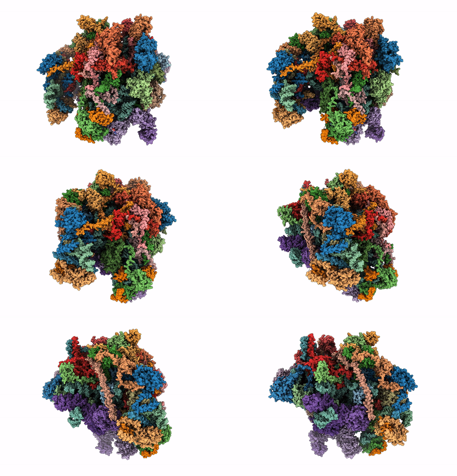 |
Mouse Mitoribosome Large Subunit Assembly Intermediate Bound To Nsun4, Metrf4, Gtpbp7, Gtpbp10 And The Malsu-L0R8F8-Mtacp Complex With Ul16M, State B2 (Samc Knock-Out)
Organism: Mus musculus
Method: ELECTRON MICROSCOPY Release Date: 2025-07-09 Classification: RIBOSOME Ligands: MG, FES, ZN, GNP |
 |
Mouse Mitoribosome Large Subunit Assembly Intermediate (Without Ul16M) Bound To Mrm3-Dimer, Ddx28 And The Malsu-L0R8F8-Mt-Acp Complex, State A1 (Samc Knock-Out)
Organism: Mus musculus
Method: ELECTRON MICROSCOPY Release Date: 2025-06-18 Classification: RIBOSOME Ligands: ZN, FES |
 |
Mouse Mitoribosome Large Subunit Assembly Intermediate (Without Ul16M) Bound To Mrm3 Dimer And The Malsu-L0R8F8-Mt-Acp Complex, State A3 (Samc Knock Out)
Organism: Mus musculus
Method: ELECTRON MICROSCOPY Release Date: 2025-06-18 Classification: RIBOSOME Ligands: ZN, FES |
 |
Mouse Mitoribosome Large Subunit Assembly Intermediate Bound To Nsun4, Metrf4, Mrm2, Gtpbp7 And Malsu1-L0R8F8-Mt-Acp Complex, State D (Samc Knock-Out)
Organism: Mus musculus
Method: ELECTRON MICROSCOPY Release Date: 2025-06-18 Classification: RIBOSOME Ligands: MG, ZN, FES, SAH |
 |
Mouse Mitoribosome Large Subunit Assembly Intermediate Bound To Nsun4, Metrf4, Gtpbp7 And The Malsu1-L0R8F8-Mt-Acp Complex, State C1 (Samc Knock-Out)
Organism: Mus musculus
Method: ELECTRON MICROSCOPY Release Date: 2025-06-18 Classification: RIBOSOME Ligands: MG, ZN, FES, GNP |
 |
Mouse Mitoribosome Large Subunit Assembly Intermediate (With Ul16M) Bound To Mrm3-Dimer, Ddx28 And The Malsu-L0R8F8-Mt-Acp Complex, State A2 (Samc Knock-Out)
Organism: Mus musculus
Method: ELECTRON MICROSCOPY Release Date: 2025-06-11 Classification: RIBOSOME Ligands: ZN, FES |
 |
The Secreted Adhesin Etpa Of Enterotoxigenic Escherichia Coli In Complex With The Mouse Mab 1C08
Organism: Escherichia coli etec h10407, Mus musculus
Method: ELECTRON MICROSCOPY Release Date: 2024-09-11 Classification: CELL ADHESION Ligands: BGC |
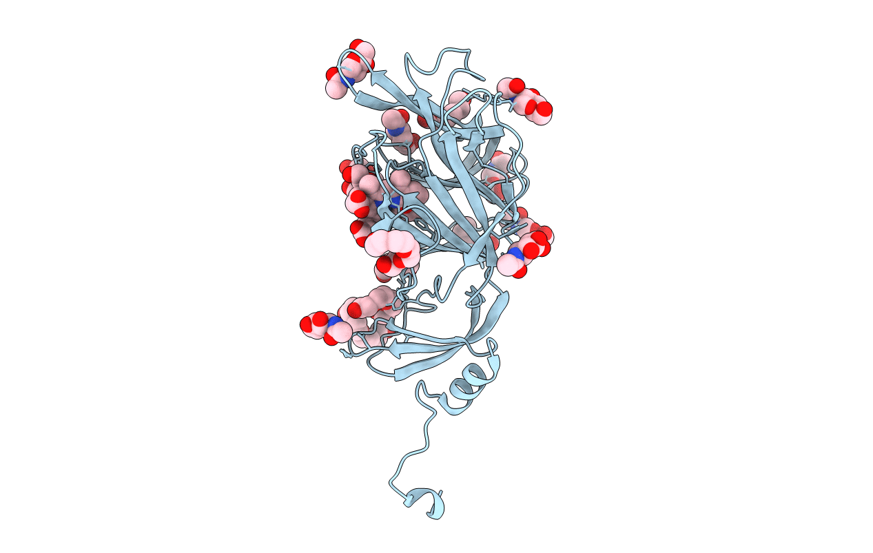 |
Crystal Structure Of Sars-Cov-2 Spike Protein N-Terminal Domain In Complex With Biliverdin
Organism: Severe acute respiratory syndrome coronavirus 2
Method: X-RAY DIFFRACTION Resolution:1.82 Å Release Date: 2021-04-28 Classification: VIRAL PROTEIN Ligands: BLA, NAG, PG4, PEG, PGE, 1PE |
 |
Trimeric Sars-Cov-2 Spike Ectodomain In Complex With Biliverdin (Closed Conformation)
Organism: Severe acute respiratory syndrome coronavirus 2
Method: ELECTRON MICROSCOPY Release Date: 2021-04-28 Classification: VIRAL PROTEIN Ligands: BLA, NAG |
 |
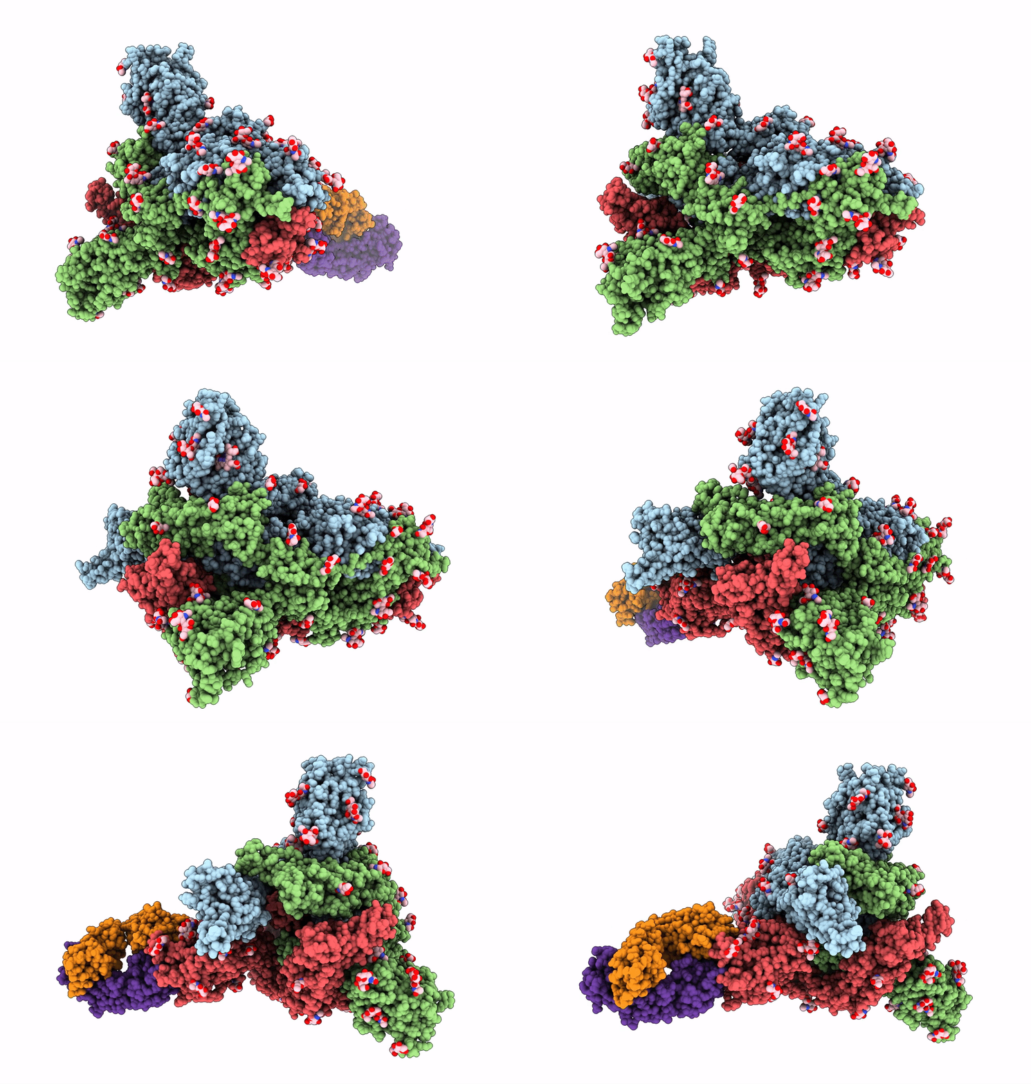
Trimeric Sars-Cov-2 Spike Ectodomain In Complex With Biliverdin (One Rbd Erect)
Organism: Severe acute respiratory syndrome coronavirus 2
Method: ELECTRON MICROSCOPY Release Date: 2021-04-28 Classification: VIRAL PROTEIN Ligands: BLA, NAG |
 |
Organism: Severe acute respiratory syndrome coronavirus 2, Homo sapiens
Method: ELECTRON MICROSCOPY Release Date: 2021-04-28 Classification: VIRAL PROTEIN Ligands: BLA, NAG |
 |
Organism: Homo sapiens
Method: X-RAY DIFFRACTION Resolution:1.90 Å Release Date: 2020-08-26 Classification: SIGNALING PROTEIN Ligands: GOL, ACT |
 |
Organism: Homo sapiens
Method: X-RAY DIFFRACTION Resolution:1.80 Å Release Date: 2020-04-01 Classification: TRANSFERASE |
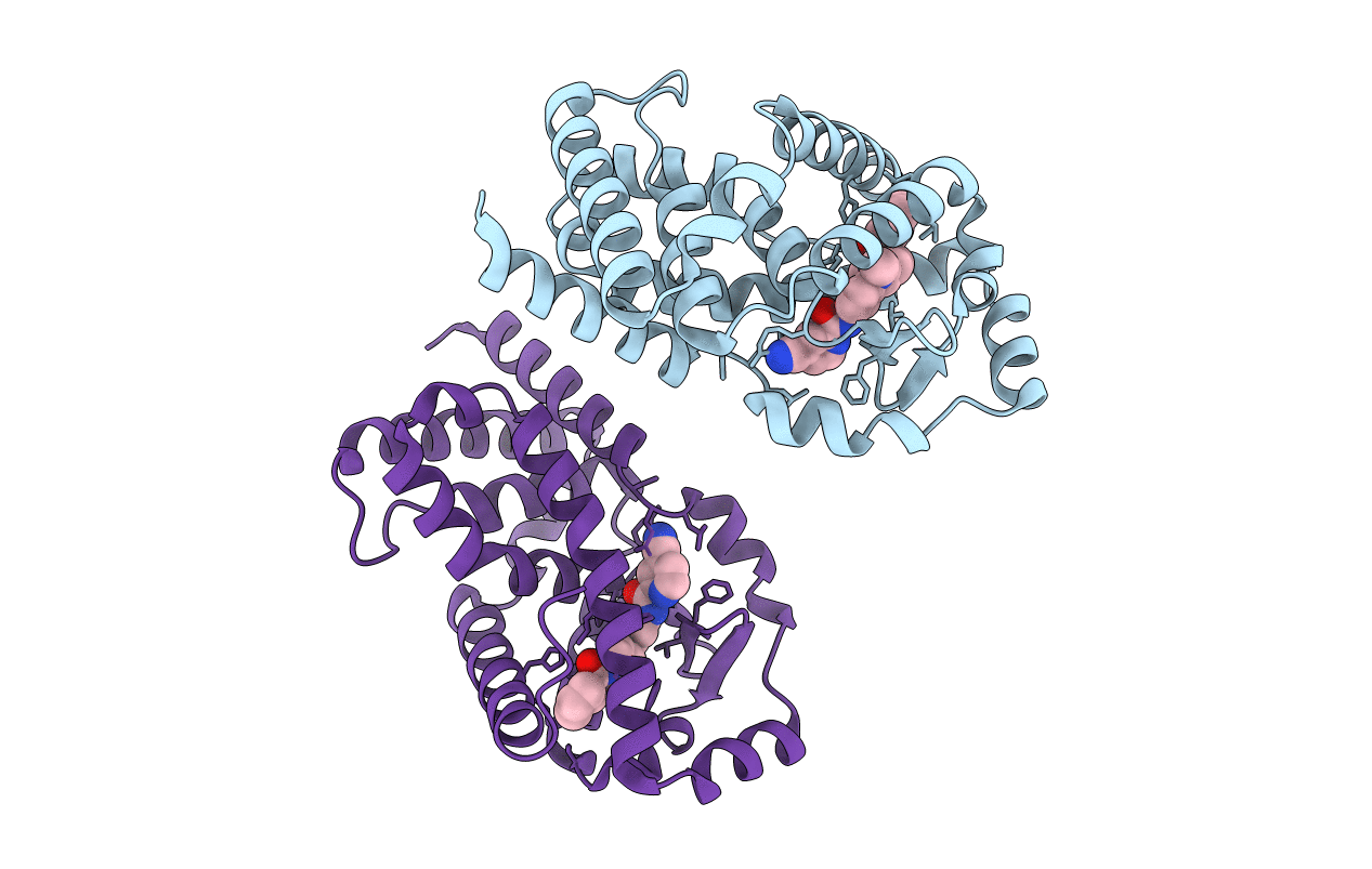 |
Human Retenoid-Related Orphan Receptor-Gamma Ligand- Binding Domain In Complex With Indole Ligand Cp9B In Inverse Agonist Conformation
Organism: Homo sapiens
Method: X-RAY DIFFRACTION Resolution:2.30 Å Release Date: 2018-09-05 Classification: SIGNALING PROTEIN Ligands: F7M |
 |
Organism: Homo sapiens
Method: X-RAY DIFFRACTION Resolution:2.45 Å Release Date: 2018-09-05 Classification: SIGNALING PROTEIN Ligands: F7J |
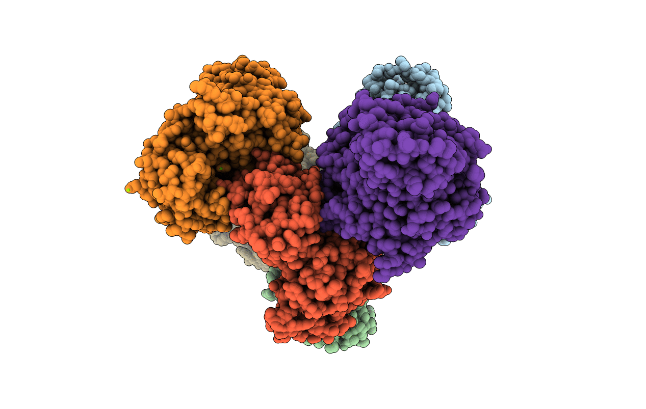 |
Organism: Tribolium castaneum
Method: X-RAY DIFFRACTION Resolution:2.78 Å Release Date: 2017-10-11 Classification: Kinase Ligands: MG |
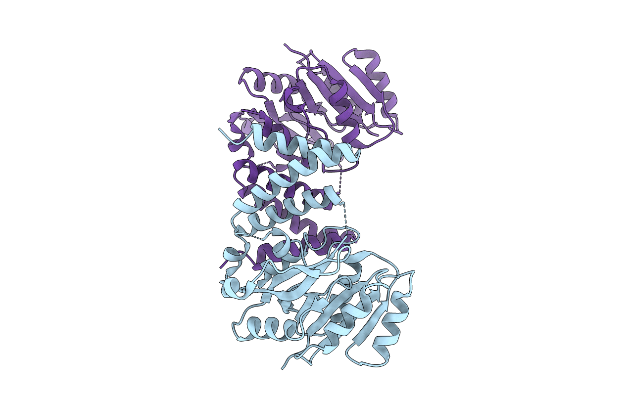 |
Organism: Streptococcus parasanguinis fw213
Method: X-RAY DIFFRACTION Resolution:2.40 Å Release Date: 2016-08-31 Classification: TRANSFERASE |
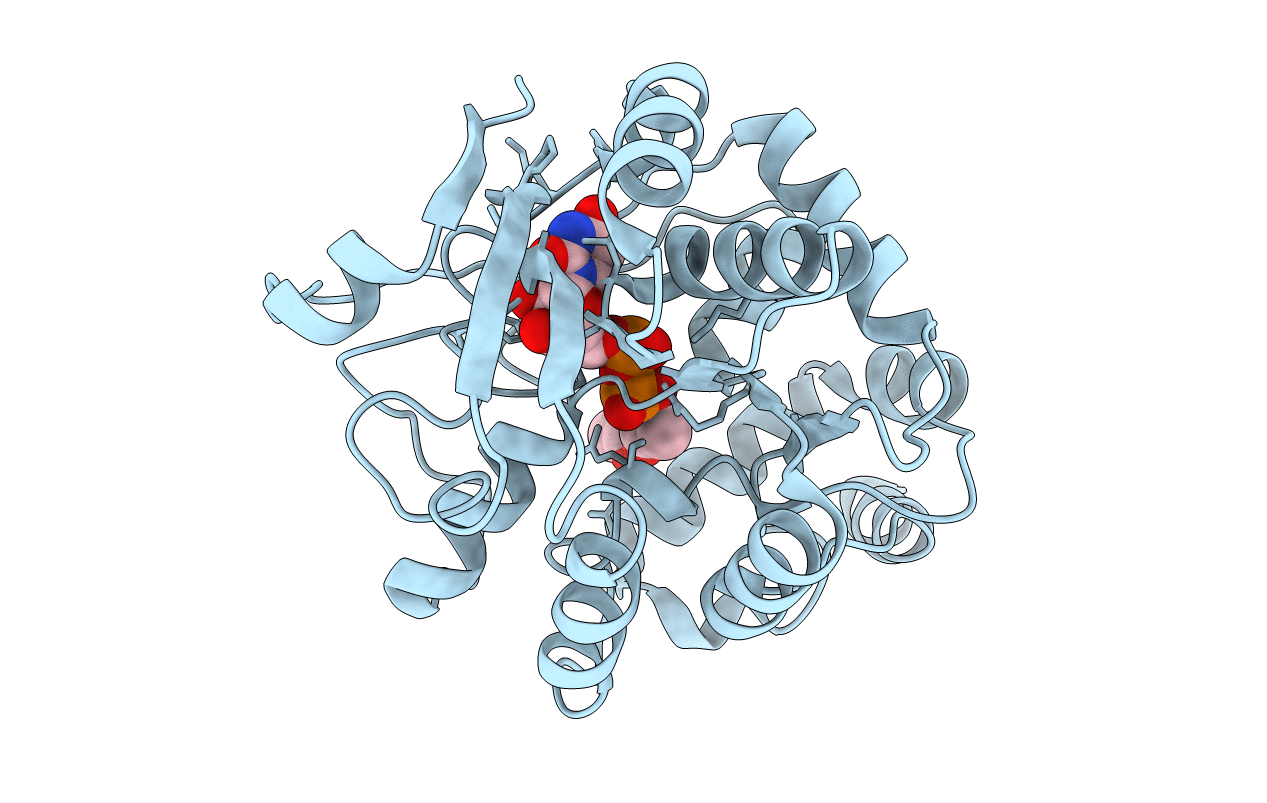 |
Organism: Streptococcus parasanguinis
Method: X-RAY DIFFRACTION Resolution:1.34 Å Release Date: 2014-08-06 Classification: TRANSFERASE Ligands: UDP, MN, ACT |
 |
Selenomethionine Substituted Structure Of Domain Of Unknown Function 1792 (Duf1792)
Organism: Streptococcus parasanguinis
Method: X-RAY DIFFRACTION Resolution:1.54 Å Release Date: 2014-08-06 Classification: TRANSFERASE Ligands: UDP |
 |
The Highly Conserved Domain Of Unknown Function 1792 Has A Distinct Glycosyltransferase Fold
Organism: Streptococcus parasanguinis
Method: X-RAY DIFFRACTION Resolution:1.66 Å Release Date: 2014-07-23 Classification: TRANSFERASE Ligands: UDP, ACT |

