
Search Count: 9
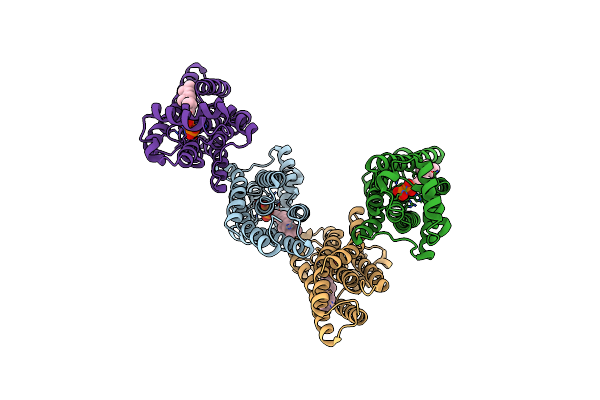 |
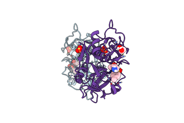
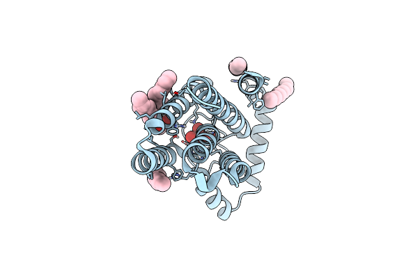
H207A Mutant Of E. Coli Pgpb, A Pap2 Type Phosphatidyl Glycerol Phosphate And C55-Pp Phosphatase, In Complex With Farnesyl Pyrophosphate
Organism: Escherichia coli k-12
Method: X-RAY DIFFRACTION Resolution:3.50 Å Release Date: 2023-11-22 Classification: HYDROLASE Ligands: FPP |
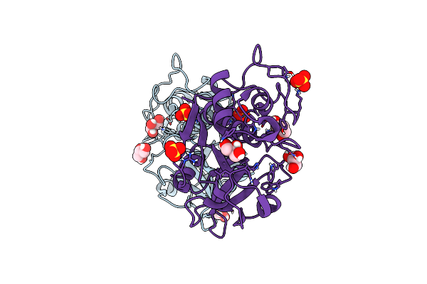 |
Structure The Ananain Protease From Ananas Comosus With A Thiomethylated Catalytic Cysteine
Organism: Ananas comosus
Method: X-RAY DIFFRACTION Resolution:1.30 Å Release Date: 2020-11-25 Classification: HYDROLASE Ligands: SO4, GOL |
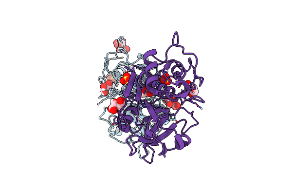 |
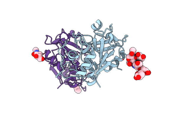
Structure The Ananain Protease From Ananas Comosus Covalently Bound To With The E64 Inhibitor
Organism: Ananas comosus
Method: X-RAY DIFFRACTION Resolution:1.26 Å Release Date: 2020-11-25 Classification: HYDROLASE Ligands: SO4, GOL, TLA |
 |
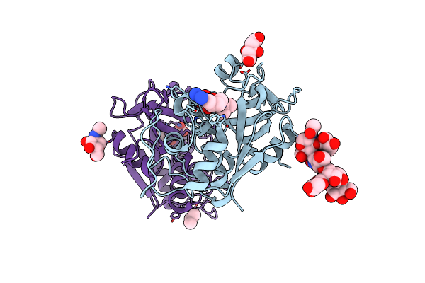
Structure The Ananain Protease From Ananas Comosus Covalently Bound To The E64 Inhibitor
Organism: Ananas comosus
Method: X-RAY DIFFRACTION Resolution:1.30 Å Release Date: 2020-11-25 Classification: HYDROLASE Ligands: E64, SO4, GOL |
 |
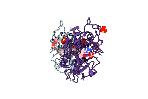
Structure The Ananain Protease From Ananas Comosus Covalently Bound To The Tlck Inhibitor
Organism: Ananas comosus
Method: X-RAY DIFFRACTION Resolution:1.35 Å Release Date: 2020-11-25 Classification: HYDROLASE Ligands: TCK, SO4, GOL |
 |
Structure The Bromelain Protease From Ananas Comosus With A Thiomethylated Active Cysteine
Organism: Ananas comosus
Method: X-RAY DIFFRACTION Resolution:1.80 Å Release Date: 2020-11-25 Classification: HYDROLASE Ligands: NAG, IPA |
 |
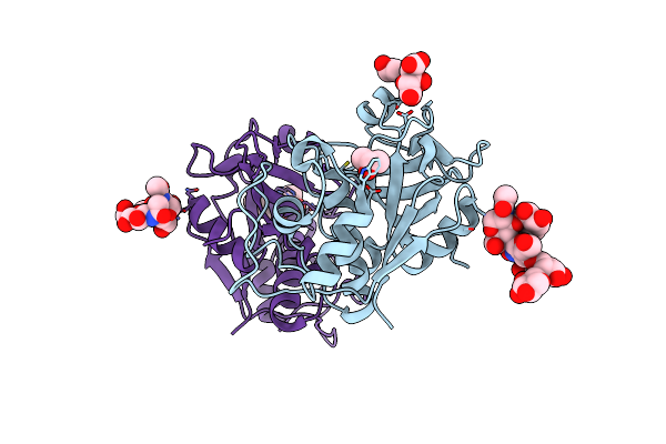
Structure The Bromelain Protease From Ananas Comosus In Complex With The E64 Inhibitor
Organism: Ananas comosus
Method: X-RAY DIFFRACTION Resolution:1.85 Å Release Date: 2020-11-25 Classification: HYDROLASE Ligands: E64, CIT, NAG, IPA |
 |
Structure The Bromelain Protease From Ananas Comosus In Complex With The Tlck Inhibitor
Organism: Ananas comosus
Method: X-RAY DIFFRACTION Resolution:1.45 Å Release Date: 2020-11-25 Classification: HYDROLASE Ligands: IPA, CIT, TCK |
 |
Crystal Structure Of The First Transmembrane Pap2 Type Phosphatidylglycerolphosphate Phosphatase From Bacillus Subtilis
Organism: Bacillus subtilis (strain 168)
Method: X-RAY DIFFRACTION Release Date: 2017-02-22 Classification: HYDROLASE Ligands: WO4, OLC, UNL |