Search Count: 45
 |
Organism: Lama glama
Method: X-RAY DIFFRACTION Resolution:2.32 Å Release Date: 2012-08-22 Classification: SIGNALING PROTEIN, HORMONE Ligands: PO4, CL |
 |
Crystal Structure Of Monomeric Isocitrate Dehydrogenase From Corynebacterium Glutamicum In Complex With Nadp
Organism: Corynebacterium glutamicum
Method: X-RAY DIFFRACTION Resolution:1.90 Å Release Date: 2011-04-06 Classification: OXIDOREDUCTASE Ligands: MG, NAP |
 |
Organism: Escherichia coli
Method: X-RAY DIFFRACTION Resolution:1.00 Å Release Date: 2008-10-21 Classification: TRANSFERASE Ligands: SO4 |
 |
Organism: Sinorhizobium meliloti
Method: X-RAY DIFFRACTION Resolution:1.95 Å Release Date: 2008-06-17 Classification: LYASE |
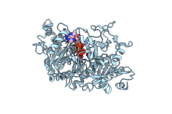 |
E. Coli Phosphoenolpyruvate Carboxykinase (Pepck) Complexed With Atp, Mg2+, Mn2+, Carbon Dioxide And Oxaloacetate
Organism: Escherichia coli
Method: X-RAY DIFFRACTION Resolution:2.23 Å Release Date: 2008-04-22 Classification: LYASE Ligands: MG, MN, OAA, ATP, CO2 |
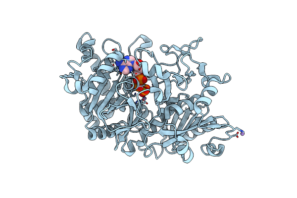 |
Crystal Structure Of E. Coli Phosphoenolpyruvate Carboxykinase Mutant Lys213Ser Complexed With Atp-Mg2+-Mn2+
Organism: Escherichia coli
Method: X-RAY DIFFRACTION Resolution:2.20 Å Release Date: 2008-04-22 Classification: LYASE Ligands: MG, MN, ATP |
 |
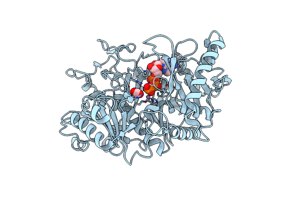
Structure Of A Gtp-Dependent Bacterial Pep-Carboxykinase From Corynebacterium Glutamicum
Organism: Corynebacterium glutamicum
Method: X-RAY DIFFRACTION Resolution:2.30 Å Release Date: 2008-04-15 Classification: SIGNALING PROTEIN, LYASE |
 |
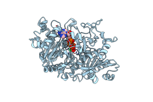
Organism: Escherichia coli
Method: X-RAY DIFFRACTION Resolution:1.94 Å Release Date: 2007-06-12 Classification: LYASE Ligands: MN, MG, ATP, CO2 |
 |
Crystal Structure Of Escherichia Coli Phosphoenolpyruvate Carboxykinase Complexed With Carbon Dioxide, Mg2+, Atp
Organism: Escherichia coli k12
Method: X-RAY DIFFRACTION Resolution:1.60 Å Release Date: 2007-06-12 Classification: LYASE Ligands: MG, CL, CO2, ATP |
 |
Organism: Staphylococcus aureus
Method: X-RAY DIFFRACTION Resolution:1.50 Å Release Date: 2007-01-02 Classification: HYDROLASE Ligands: K |
 |
Organism: Pyrobaculum aerophilum
Method: X-RAY DIFFRACTION Resolution:2.25 Å Release Date: 2006-06-06 Classification: TRANSFERASE Ligands: ADN, PO4 |
 |
Organism: Corynebacterium glutamicum
Method: X-RAY DIFFRACTION Resolution:1.75 Å Release Date: 2006-01-31 Classification: OXIDOREDUCTASE Ligands: MG |
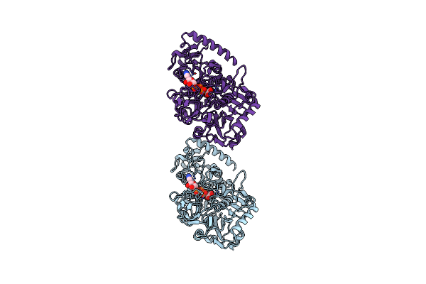 |
Crystal Structure Of Phosphoenolpyruvate Carboxykinase Of Anaerobiospirillum Succiniciproducens Complexed With Atp, Oxalate, Magnesium And Manganese Ions
Organism: Anaerobiospirillum succiniciproducens
Method: X-RAY DIFFRACTION Resolution:2.20 Å Release Date: 2006-01-24 Classification: LYASE Ligands: MG, MN, ATP, OXD |
 |
Structure Of Conserved Hypothetical Protein Pae2307 From Pyrobaculum Aerophilum
Organism: Pyrobaculum aerophilum
Method: X-RAY DIFFRACTION Resolution:1.45 Å Release Date: 2006-01-10 Classification: STRUCTURAL GENOMICS, UNKNOWN FUNCTION Ligands: PO4 |
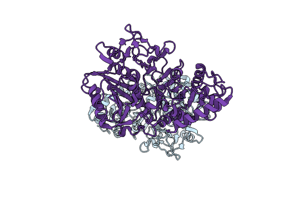 |
Crystal Structure Of Anaerobiospirillum Succiniciproducens Phosphoenolpyruvate Carboxykinase
Organism: Anaerobiospirillum succiniciproducens
Method: X-RAY DIFFRACTION Resolution:2.35 Å Release Date: 2005-07-26 Classification: LYASE |
 |
Crystal Structure Of Phosphoenolpyruvate Carboxykinase From Actinobacillus Succinogenes
Organism: Actinobacillus succinogenes
Method: X-RAY DIFFRACTION Resolution:1.85 Å Release Date: 2005-06-28 Classification: LYASE Ligands: SO4, NA |
 |
Crystal Structure Of Phosphoenolpyruvate Carboxykinase From Actinobaccilus Succinogenes In Complex With Manganese And Pyruvate
Organism: Actinobacillus succinogenes
Method: X-RAY DIFFRACTION Resolution:1.70 Å Release Date: 2005-06-28 Classification: LYASE Ligands: MN, PO4, DT3, BME, PYR, FMT |
 |
Organism: Staphylococcus aureus subsp. aureus mu50
Method: X-RAY DIFFRACTION Resolution:1.90 Å Release Date: 2004-02-17 Classification: Protease Ligands: K |
 |
Method: X-RAY DIFFRACTION
Resolution:3.02 Å Release Date: 2003-12-09 Classification: DNA Ligands: CO |
 |
Method: X-RAY DIFFRACTION
Resolution:2.90 Å Release Date: 2003-12-09 Classification: DNA Ligands: NI |

