
Search Count: 291
 |
Organism: Mycosarcoma maydis
Method: X-RAY DIFFRACTION Release Date: 2025-08-20 Classification: UNKNOWN FUNCTION Ligands: SO4 |
 |
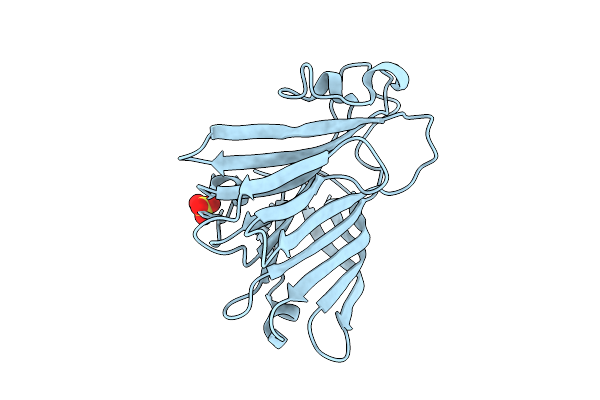
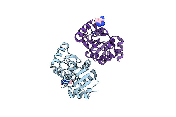
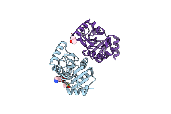
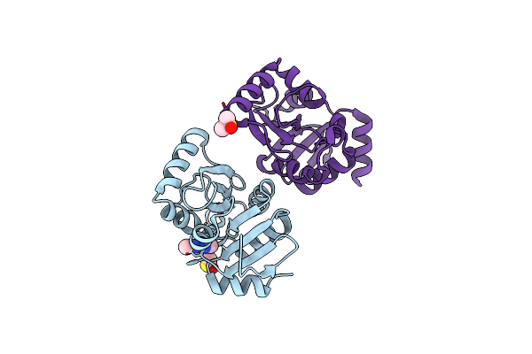
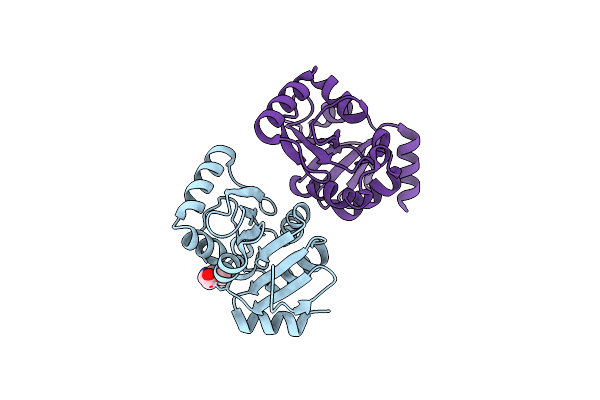
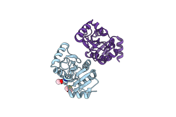
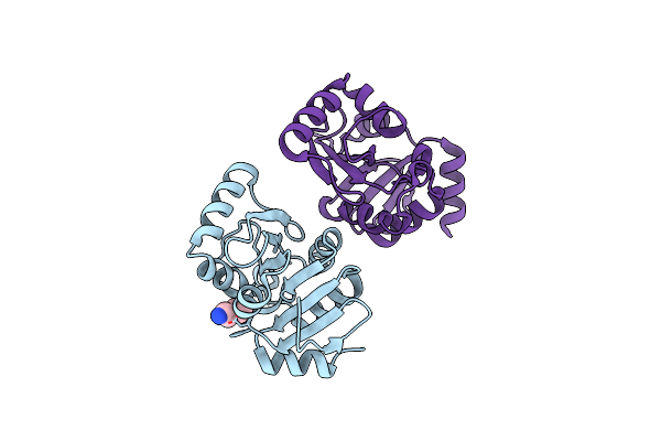
Organism: Phytophthora infestans
Method: X-RAY DIFFRACTION Resolution:2.30 Å Release Date: 2025-01-15 Classification: HYDROLASE |
 |
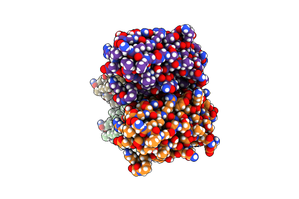
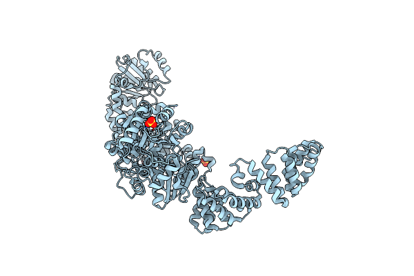
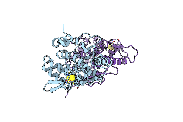
Organism: Ustilago maydis 521
Method: X-RAY DIFFRACTION Resolution:1.81 Å Release Date: 2025-01-15 Classification: HYDROLASE |
 |
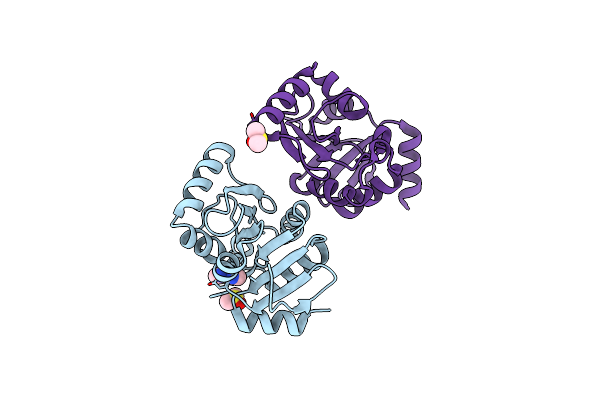
Organism: Mus musculus, Synthetic construct
Method: ELECTRON MICROSCOPY Release Date: 2024-10-16 Classification: MEMBRANE PROTEIN |
 |
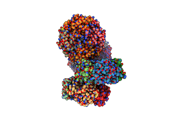
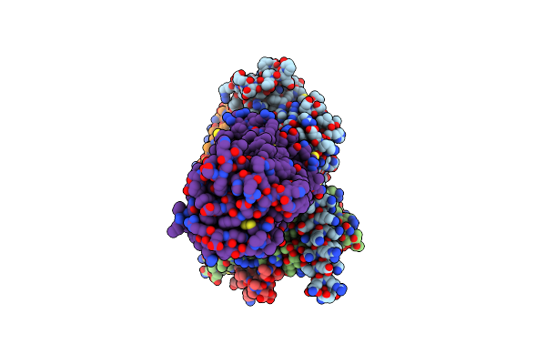
Capsular Polysaccharide Synthesis Multienzyme Of Actinobacillus Pleuropneumoniae Serotype 3
Organism: Actinobacillus pleuropneumoniae
Method: X-RAY DIFFRACTION Resolution:3.00 Å Release Date: 2024-07-03 Classification: BIOSYNTHETIC PROTEIN Ligands: SO4, ZN |
 |
Organism: Homo sapiens, Lama glama
Method: ELECTRON MICROSCOPY Release Date: 2024-02-21 Classification: MEMBRANE PROTEIN Ligands: CLR |
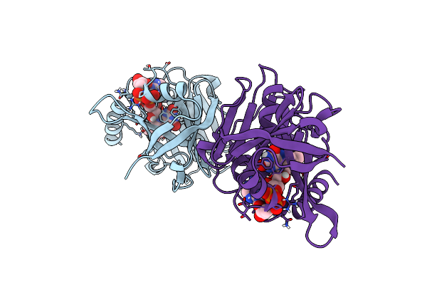 |
Ferric-Siderophore Reduction In Shewanella Biscestrii: Structural And Functional Characterization Of Sbisip Reveals Unforeseen Specificity
Organism: Shewanella bicestrii
Method: X-RAY DIFFRACTION Release Date: 2024-01-17 Classification: METAL BINDING PROTEIN Ligands: FAD |
 |
Organism: Juglans regia
Method: X-RAY DIFFRACTION Resolution:3.10 Å Release Date: 2023-12-20 Classification: PROTEIN BINDING |
 |
Organism: Escherichia coli k-12
Method: X-RAY DIFFRACTION Resolution:1.92 Å Release Date: 2023-07-12 Classification: OXIDOREDUCTASE Ligands: FES |
 |
Organism: Haemophilus influenzae
Method: X-RAY DIFFRACTION Resolution:2.90 Å Release Date: 2023-04-26 Classification: BIOSYNTHETIC PROTEIN Ligands: PO4, GOL, ZN, C5P |
 |
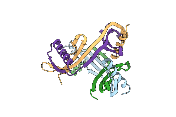
Capsular Polysaccharide Synthesis Multienzyme In Complex With Capsular Polymer Fragment
Organism: Haemophilus influenzae
Method: X-RAY DIFFRACTION Resolution:3.60 Å Release Date: 2023-04-26 Classification: BIOSYNTHETIC PROTEIN Ligands: MG, KOF |
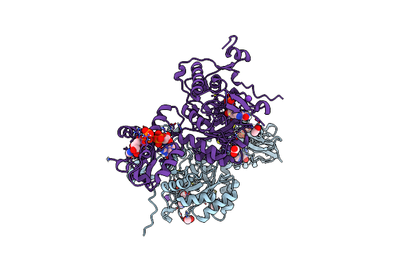 |
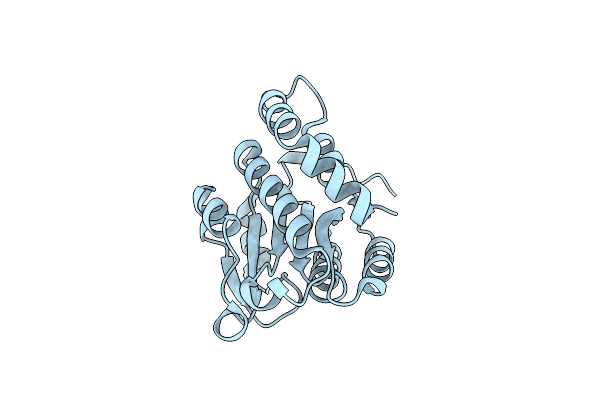
Crystal Structure Of Pseudomonas Aeruginosa Guab (Imp Dehydrogenase) Bound To Atp And Gdp At 1.65A Resolution
Organism: Pseudomonas aeruginosa
Method: X-RAY DIFFRACTION Resolution:1.65 Å Release Date: 2022-07-06 Classification: OXIDOREDUCTASE Ligands: ATP, GDP, IMP, MG, K, ACT |
 |
Crystal Structure Of Streptomyces Coelicolor Guab (Imp Dehydrogenase) Bound To Atp And Ppgpp At 2.0 A Resolution
Organism: Streptomyces coelicolor a3(2)
Method: X-RAY DIFFRACTION Resolution:2.03 Å Release Date: 2022-05-11 Classification: OXIDOREDUCTASE Ligands: ATP, G4P, MG |
 |
Pandda Analysis Group Deposition -- Crystal Structure Of Sars-Cov-2 Nsp3 Macrodomain In Complex With En300-321461
Organism: Severe acute respiratory syndrome coronavirus 2
Method: X-RAY DIFFRACTION Resolution:1.13 Å Release Date: 2021-01-13 Classification: VIRAL PROTEIN Ligands: WOY, DMS |
 |
Pandda Analysis Group Deposition -- Crystal Structure Of Sars-Cov-2 Nsp3 Macrodomain In Complex With En300-43406
Organism: Severe acute respiratory syndrome coronavirus 2
Method: X-RAY DIFFRACTION Resolution:1.08 Å Release Date: 2021-01-13 Classification: VIRAL PROTEIN Ligands: WPS |
 |
Pandda Analysis Group Deposition -- Crystal Structure Of Sars-Cov-2 Nsp3 Macrodomain In Complex With Z3034471507
Organism: Severe acute respiratory syndrome coronavirus 2
Method: X-RAY DIFFRACTION Resolution:1.17 Å Release Date: 2021-01-13 Classification: VIRAL PROTEIN Ligands: WPV, DMS |
 |
Pandda Analysis Group Deposition -- Crystal Structure Of Sars-Cov-2 Nsp3 Macrodomain In Complex With Ab-601_30915014
Organism: Severe acute respiratory syndrome coronavirus 2
Method: X-RAY DIFFRACTION Resolution:1.17 Å Release Date: 2021-01-13 Classification: VIRAL PROTEIN Ligands: WPY, DMS |
 |
Pandda Analysis Group Deposition -- Crystal Structure Of Sars-Cov-2 Nsp3 Macrodomain In Complex With En300-108952
Organism: Severe acute respiratory syndrome coronavirus 2
Method: X-RAY DIFFRACTION Resolution:1.11 Å Release Date: 2021-01-13 Classification: VIRAL PROTEIN Ligands: WQ1 |
 |
Pandda Analysis Group Deposition -- Crystal Structure Of Sars-Cov-2 Nsp3 Macrodomain In Complex With En300-301084
Organism: Severe acute respiratory syndrome coronavirus 2
Method: X-RAY DIFFRACTION Resolution:1.07 Å Release Date: 2021-01-13 Classification: VIRAL PROTEIN Ligands: WQ4, DMS |
 |
Pandda Analysis Group Deposition -- Crystal Structure Of Sars-Cov-2 Nsp3 Macrodomain In Complex With En300-105873
Organism: Severe acute respiratory syndrome coronavirus 2
Method: X-RAY DIFFRACTION Resolution:1.08 Å Release Date: 2021-01-13 Classification: VIRAL PROTEIN Ligands: WQ7 |