
Search Count: 17
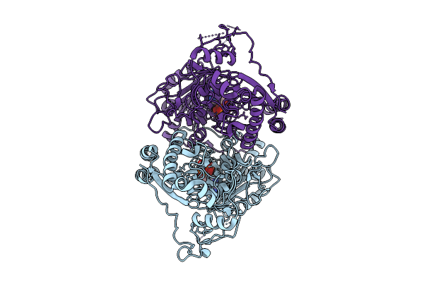 |
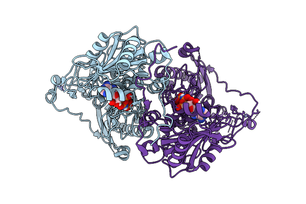
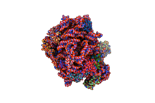
Organism: Homo sapiens
Method: ELECTRON MICROSCOPY Release Date: 2025-07-09 Classification: DNA BINDING PROTEIN Ligands: ZN, ANP, MG |
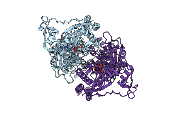 |
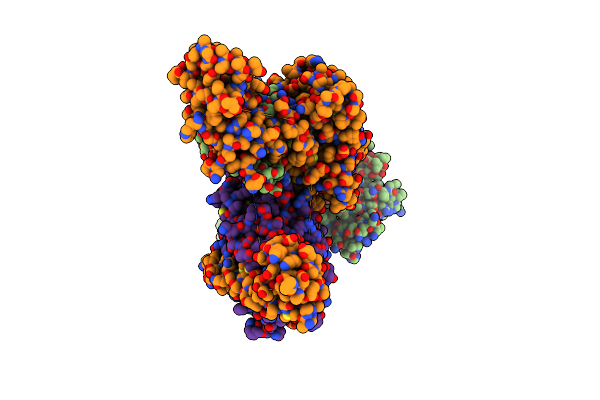
Organism: Homo sapiens
Method: ELECTRON MICROSCOPY Release Date: 2025-07-09 Classification: DNA BINDING PROTEIN Ligands: ZN, ATP, MG |
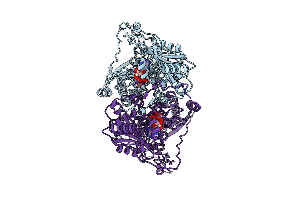 |
Organism: Homo sapiens
Method: ELECTRON MICROSCOPY Release Date: 2025-07-09 Classification: DNA BINDING PROTEIN Ligands: ZN, ANP, MG |
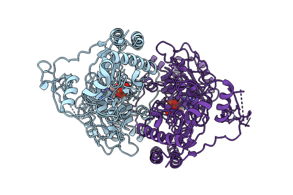 |
Organism: Homo sapiens
Method: ELECTRON MICROSCOPY Release Date: 2025-07-09 Classification: DNA BINDING PROTEIN Ligands: ZN, ANP, MG |
 |
Organism: Homo sapiens
Method: ELECTRON MICROSCOPY Release Date: 2025-07-09 Classification: DNA BINDING PROTEIN Ligands: ZN, ANP, MG |
 |
Organism: Homo sapiens
Method: ELECTRON MICROSCOPY Release Date: 2024-07-31 Classification: GENE REGULATION |
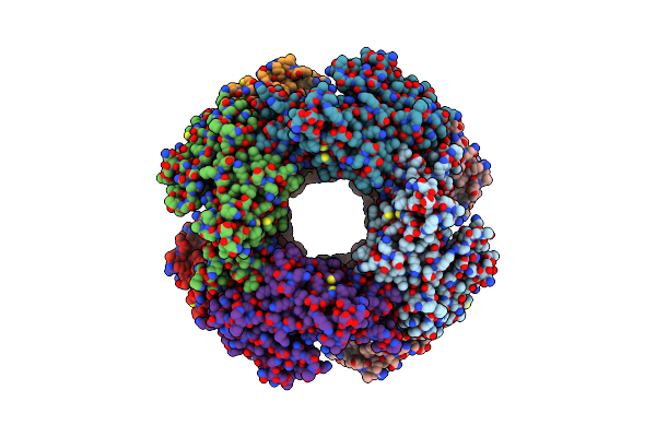 |
Organism: Rotavirus a
Method: ELECTRON MICROSCOPY Release Date: 2021-09-29 Classification: VIRAL PROTEIN |
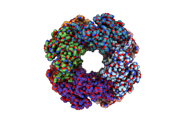 |
Organism: Rotavirus a
Method: ELECTRON MICROSCOPY Release Date: 2021-09-29 Classification: VIRAL PROTEIN |
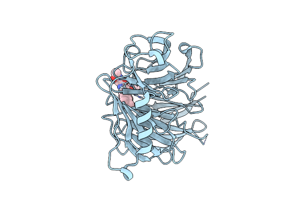 |
Organism: Homo sapiens
Method: X-RAY DIFFRACTION Resolution:1.70 Å Release Date: 2021-05-26 Classification: GENE REGULATION |
 |
Organism: Homo sapiens
Method: X-RAY DIFFRACTION Resolution:1.63 Å Release Date: 2021-05-26 Classification: GENE REGULATION |
 |
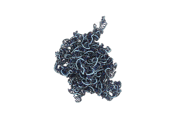
Crystal Structure Of The Large Ribosomal Subunit (50S) Of Deinococcus Radiodurans Containing A Three Residue Insertion In L22 In Complex With Erythromycin
Organism: Deinococcus radiodurans (strain atcc 13939 / dsm 20539 / jcm 16871 / lmg 4051 / nbrc 15346 / ncimb 9279 / r1 / vkm b-1422), Deinococcus radiodurans, Deinococcus radiodurans r1
Method: X-RAY DIFFRACTION Resolution:3.54 Å Release Date: 2015-12-23 Classification: RIBOSOME Ligands: MG, ERY |
 |
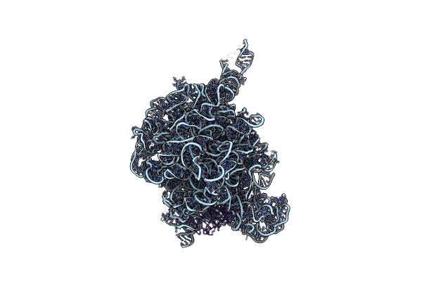
Crystal Structure Of The Large Ribosomal Subunit (50S) Of Deinococcus Radiodurans Containing A Three Residue Insertion In L22
Organism: Deinococcus radiodurans
Method: X-RAY DIFFRACTION Resolution:3.65 Å Release Date: 2015-08-05 Classification: RIBOSOME Ligands: MG |
 |
The Structure Of The Complex Of The Large Ribosomal Subunit From D. Radiodurans With The Antibiotic Lankacidin
Organism: Deinococcus radiodurans
Method: X-RAY DIFFRACTION Resolution:3.52 Å Release Date: 2010-09-08 Classification: RNA Ligands: LC2 |
 |
The Large Ribosomal Subunit From Deinococcus Radiodurans Complexed With Methymycin
Organism: Deinococcus radiodurans r1
Method: X-RAY DIFFRACTION Resolution:3.71 Å Release Date: 2010-01-19 Classification: RIBOSOME Ligands: MT9 |
 |
The Crystal Structure Of The Large Ribosomal Subunit From Deinococcus Radiodurans Complexed With The Pleuromutilin Derivative Sb-571519
Organism: Deinococcus radiodurans
Method: X-RAY DIFFRACTION Resolution:3.50 Å Release Date: 2007-05-01 Classification: RIBOSOME Ligands: G19 |
 |
The Crystal Structure Of The Large Ribosomal Subunit From Deinococcus Radiodurans Complexed With The Pleuromutilin Derivative Sb-280080
Organism: Deinococcus radiodurans
Method: X-RAY DIFFRACTION Resolution:3.56 Å Release Date: 2007-05-01 Classification: RIBOSOME Ligands: G80 |
 |
The Crystal Structure Of The Large Ribosomal Subunit From Deinococcus Radiodurans Complexed With The Pleuromutilin Derivative Retapamulin (Sb-275833)
Organism: Deinococcus radiodurans
Method: X-RAY DIFFRACTION Resolution:3.66 Å Release Date: 2007-05-01 Classification: RIBOSOME Ligands: G34 |