
Search Count: 33
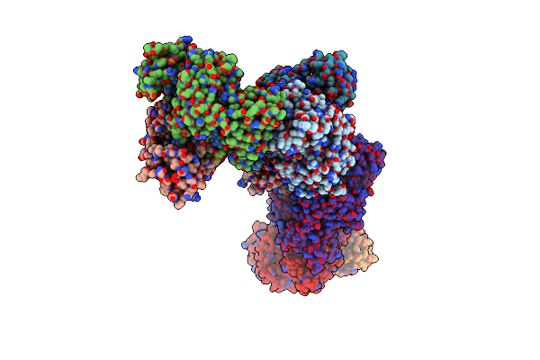 |
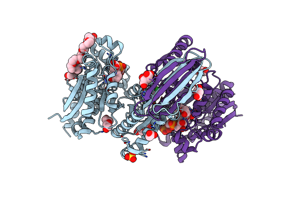
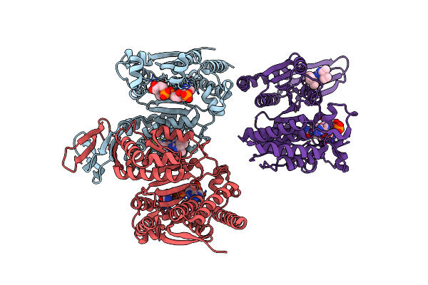
Structure Of Human Erythrocyte Pyruvate Kinase In Complex With An Allosteric Activator Ag-946
Organism: Homo sapiens
Method: X-RAY DIFFRACTION Resolution:2.35 Å Release Date: 2023-12-27 Classification: TRANSFERASE/ACTIVATOR Ligands: FBP, MN, K, PYR, HVI |
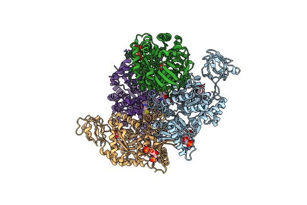 |
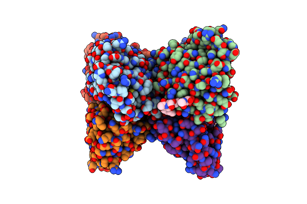
Structure Of Human Erythrocyte Pyruvate Kinase In Complex With An Allosteric Activator Compound 2
Organism: Homo sapiens
Method: X-RAY DIFFRACTION Resolution:2.34 Å Release Date: 2023-12-27 Classification: TRANSFERASE/ACTIVATOR Ligands: FBP, PYR, MN, K, I07 |
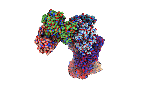 |
Structure Of Human Erythrocyte Pyruvate Kinase In Complex With An Allosteric Activator Compound 12
Organism: Homo sapiens
Method: X-RAY DIFFRACTION Resolution:2.35 Å Release Date: 2023-12-27 Classification: TRANSFERASE/ACTIVATOR Ligands: I0R, PYR, FBP, MN, K |
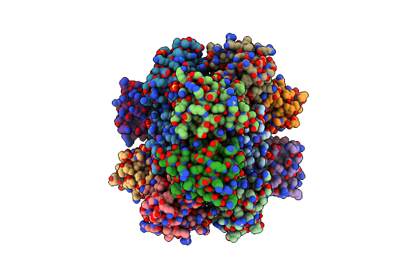 |
Organism: Human immunodeficiency virus type 2 subtype a (isolate ben)
Method: X-RAY DIFFRACTION Resolution:2.38 Å Release Date: 2023-09-13 Classification: VIRAL PROTEIN Ligands: SO4 |
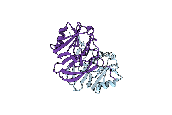 |
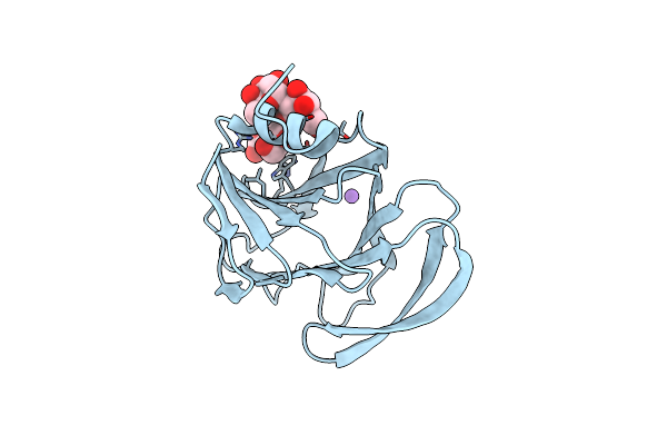
Structural And Functional Characterization Of Bovine G1P[5] Rotavirus Vp8* Protein
Organism: Rotavirus a
Method: X-RAY DIFFRACTION Resolution:1.70 Å Release Date: 2021-12-29 Classification: VIRAL PROTEIN |
 |
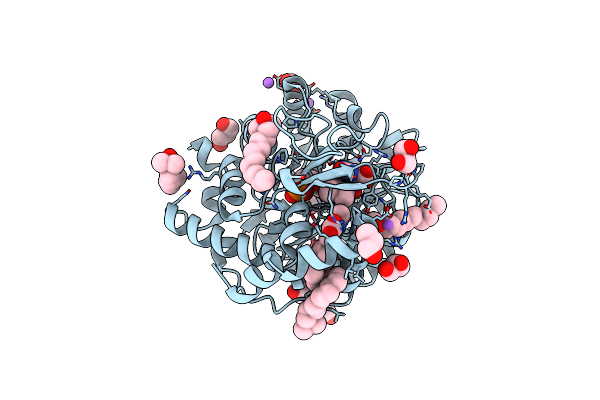
Crystal Structure Of Human Methionine Adenosyltransferase 2A (Mat2A) In Complex With Sam And Allosteric Inhibitor Ag-270
Organism: Homo sapiens
Method: X-RAY DIFFRACTION Resolution:1.32 Å Release Date: 2021-04-21 Classification: TRANSFERASE/Inhibitor Ligands: CL, EDO, SAM, WBG |
 |
Crystal Structure Of Human Methionine Adenosyltransferase 2A (Mat2A) In Complex With Sam And Allosteric Inhibitor Compound 2
Organism: Homo sapiens
Method: X-RAY DIFFRACTION Resolution:1.14 Å Release Date: 2021-04-21 Classification: TRANSFERASE/Inhibitor Ligands: J41, SAM, CL |
 |
Crystal Structure Of Human Methionine Adenosyltransferase 2A (Mat2A) In Complex With Sam And Allosteric Inhibitor Agi-24512
Organism: Homo sapiens
Method: X-RAY DIFFRACTION Resolution:1.10 Å Release Date: 2021-04-21 Classification: TRANSFERASE/Inhibitor Ligands: J4A, SAM, EDO, GOL |
 |
Crystal Structure Of Human Methionine Adenosyltransferase 2A (Mat2A) In Complex With Sam And Allosteric Inhibitor Compound 34
Organism: Homo sapiens
Method: X-RAY DIFFRACTION Resolution:1.24 Å Release Date: 2021-04-21 Classification: TRANSFERASE/Inhibitor Ligands: SAM, CL, TRS, GOL, WBM |
 |
Crystal Structure Of Human Methionine Adenosyltransferase 2A (Mat2A) In Complex With Sam And Allosteric Inhibitor Compound 35
Organism: Homo sapiens
Method: X-RAY DIFFRACTION Resolution:1.24 Å Release Date: 2021-04-21 Classification: TRANSFERASE/Inhibitor Ligands: WBS, CL, TRS, EDO, GOL, SAM |
 |
Quaternary Complex Of Human Dihydroorotate Dehydrogenase (Dhodh) With Flavin Mononucleotide (Fmn), Orotic Acid And Ag-636
Organism: Homo sapiens
Method: X-RAY DIFFRACTION Resolution:1.97 Å Release Date: 2020-11-04 Classification: OXIDOREDUCTASE/OXIDOREDUCTASE INHIBITOR Ligands: R4P, DET, DDQ, CL, NA, FMN, ORO, ACT, GOL |
 |
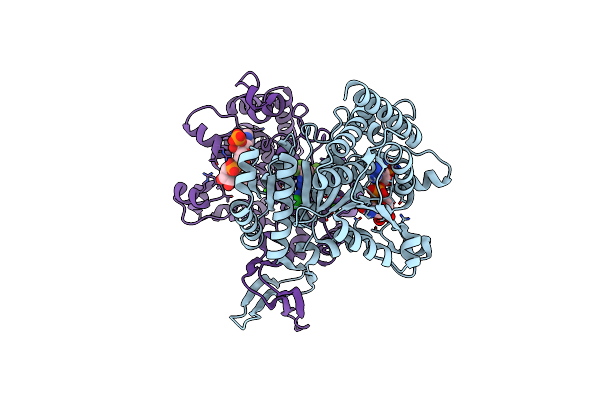
Crystal Structure Of Human Cytosolic Isocitrate Dehydrogenase (Idh1) R132H Mutant In Complex With Nadph And Ag-881 (Vorasidenib) Inhibitor
Organism: Homo sapiens
Method: X-RAY DIFFRACTION Resolution:2.10 Å Release Date: 2020-02-05 Classification: ANTITUMOR PROTEIN Ligands: NDP, SO4, 33O, PEG, ACT, MLA, 9UO, MLT |
 |
Crystal Structure Of Human Mitochondrial Isocitrate Dehydrogenase (Idh2) R140Q Mutant Homodimer In Complex With Nadph And Ag-881 (Vorasidenib) Inhibitor.
Organism: Homo sapiens
Method: X-RAY DIFFRACTION Resolution:1.99 Å Release Date: 2020-02-05 Classification: OXIDOREDUCTASE/OXIDOREDUCTASE inhibitor Ligands: 9UO, NDP, CA, NA |
 |
Crystal Structure Of Human Cytosolic Isocitrate Dehydrogenase (Idh1) R132H Mutant In Complex With Nadph And Agi-15056
Organism: Homo sapiens
Method: X-RAY DIFFRACTION Resolution:2.66 Å Release Date: 2020-02-05 Classification: OXIDOREDUCTASE/OXIDOREDUCTASE inhibitor Ligands: NDP, QWM |
 |
Structural Basis Of Glycan Recognition In Globally Predominant Human P[8] Rotavirus
Organism: Rotavirus a
Method: X-RAY DIFFRACTION Resolution:1.80 Å Release Date: 2019-10-09 Classification: VIRAL PROTEIN |
 |
Structural Basis Of Glycan Recognition In Globally Predominant Human P[8] Rotavirus
Organism: Rotavirus a
Method: X-RAY DIFFRACTION Resolution:2.30 Å Release Date: 2019-10-09 Classification: VIRAL PROTEIN |
 |
Organism: Homo sapiens, Escherichia coli
Method: X-RAY DIFFRACTION Resolution:2.26 Å Release Date: 2019-05-15 Classification: BIOSYNTHETIC PROTEIN/SIGNALING PROTEIN Ligands: MPD, ACT, ACY |
 |
Organism: Homo sapiens, Neosartorya fumigata (strain atcc mya-4609 / af293 / cbs 101355 / fgsc a1100)
Method: X-RAY DIFFRACTION Resolution:3.01 Å Release Date: 2019-05-15 Classification: BIOSYNTHETIC PROTEIN/SIGNALING PROTEIN Ligands: NAG |
 |
Organism: Escherichia coli, Homo sapiens
Method: X-RAY DIFFRACTION Resolution:1.30 Å Release Date: 2019-05-15 Classification: BIOSYNTHETIC PROTEIN/SIGNALING PROTEIN Ligands: ACT, SO4, EDO, NAG |
 |
Organism: Escherichia coli, Homo sapiens
Method: X-RAY DIFFRACTION Resolution:1.65 Å Release Date: 2019-05-15 Classification: BIOSYNTHETIC PROTEIN/SIGNALING PROTEIN Ligands: SO4, EDO |