
Search Count: 208
 |
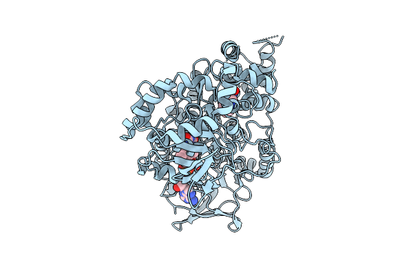
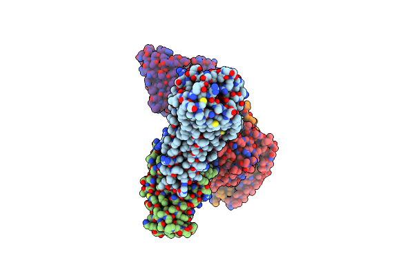
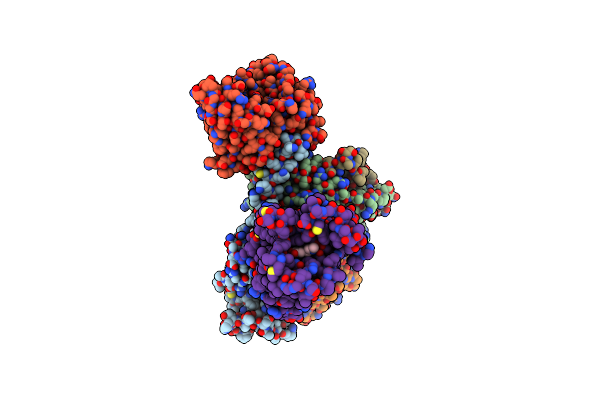
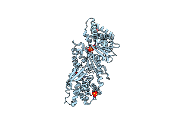
Organism: Aetokthonos hydrillicola thurmond2011
Method: X-RAY DIFFRACTION Release Date: 2025-12-10 Classification: FLAVOPROTEIN Ligands: FAD, TRP |
 |
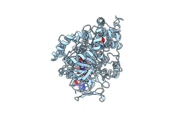
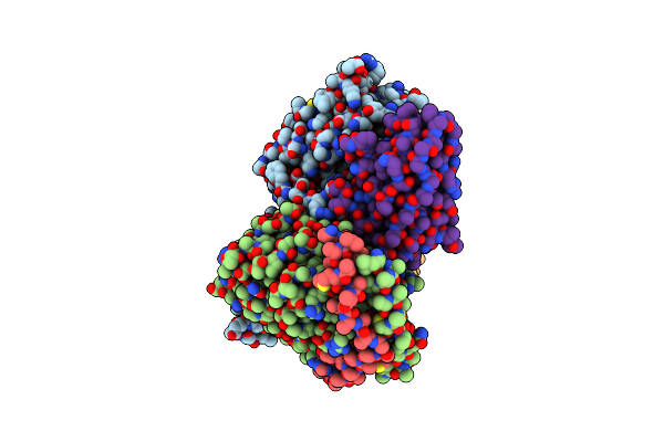
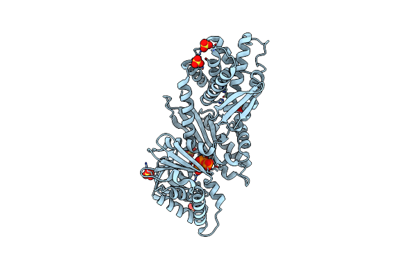
Organism: Aetokthonos hydrillicola thurmond2011
Method: X-RAY DIFFRACTION Release Date: 2025-12-10 Classification: FLAVOPROTEIN Ligands: FAD, TRP |
 |
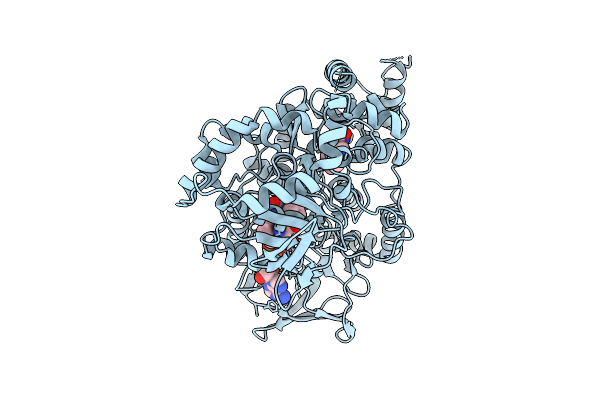
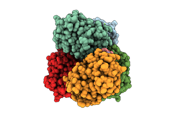
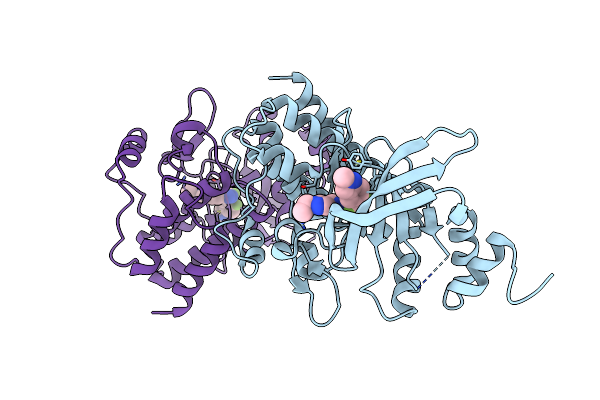
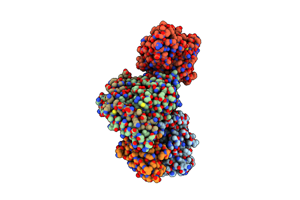
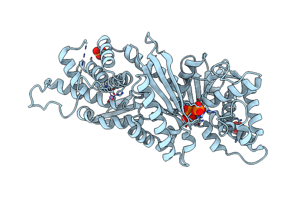
Crystal Structure Of Aetf-L183F/V220I/S523A In Complex With Fad And L-Tryptophan
Organism: Aetokthonos hydrillicola thurmond2011
Method: X-RAY DIFFRACTION Release Date: 2025-12-10 Classification: FLAVOPROTEIN Ligands: TRP, FAD |
 |
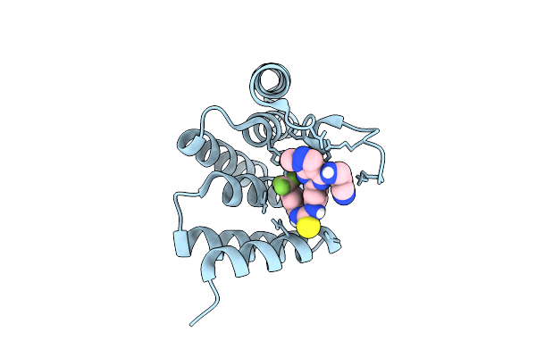
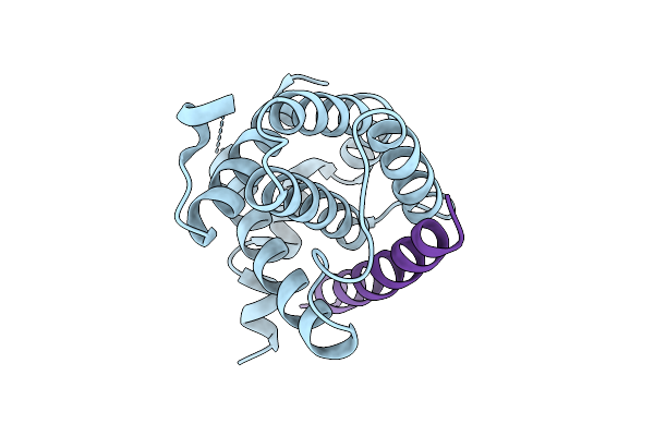
Organism: Arabidopsis thaliana
Method: X-RAY DIFFRACTION Release Date: 2025-10-29 Classification: PLANT PROTEIN Ligands: OT5 |
 |
Organism: Homo sapiens
Method: X-RAY DIFFRACTION Release Date: 2025-09-17 Classification: CELL CYCLE Ligands: A1JBJ |
 |
Organism: Homo sapiens
Method: X-RAY DIFFRACTION Release Date: 2025-08-06 Classification: APOPTOSIS Ligands: MG |
 |
 |
Organism: Homo sapiens, Vicugna pacos
Method: ELECTRON MICROSCOPY Release Date: 2025-06-11 Classification: MEMBRANE PROTEIN Ligands: UTP |
 |
Organism: Homo sapiens, Vicugna pacos
Method: ELECTRON MICROSCOPY Release Date: 2025-06-11 Classification: MEMBRANE PROTEIN Ligands: ATP |
 |
Organism: Homo sapiens, Mus musculus
Method: ELECTRON MICROSCOPY Release Date: 2025-06-11 Classification: MEMBRANE PROTEIN Ligands: ATP |
 |
Organism: Homo sapiens, Vicugna pacos
Method: ELECTRON MICROSCOPY Release Date: 2025-06-11 Classification: MEMBRANE PROTEIN |
 |
Organism: Homo sapiens
Method: X-RAY DIFFRACTION Resolution:2.35 Å Release Date: 2025-01-22 Classification: TRANSFERASE Ligands: A1L3A |
 |
Organism: Fusarium tricinctum
Method: X-RAY DIFFRACTION Resolution:2.27 Å Release Date: 2024-12-11 Classification: TRANSFERASE Ligands: PPV, TOE, MOE |
 |
Organism: Rattus norvegicus, Lama glama, Homo sapiens, Bos taurus
Method: ELECTRON MICROSCOPY Release Date: 2024-08-21 Classification: MEMBRANE PROTEIN/IMMUNE SYSTEM Ligands: CAU |
 |
Organism: Rattus norvegicus, Lama glama, Homo sapiens, Bos taurus
Method: ELECTRON MICROSCOPY Release Date: 2024-08-21 Classification: MEMBRANE PROTEIN/IMMUNE SYSTEM Ligands: ALE |
 |
Organism: Homo sapiens
Method: X-RAY DIFFRACTION Resolution:2.67 Å Release Date: 2024-07-31 Classification: TRANSFERASE Ligands: IYC |
 |
Organism: Gallus gallus
Method: X-RAY DIFFRACTION Resolution:2.80 Å Release Date: 2024-07-31 Classification: TRANSFERASE Ligands: IYC |
 |
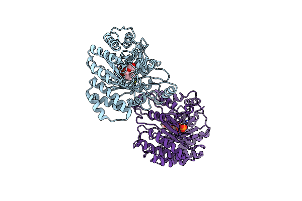
Organism: Deinococcus radiodurans r1 = atcc 13939 = dsm 20539
Method: X-RAY DIFFRACTION Resolution:1.90 Å Release Date: 2024-05-15 Classification: HYDROLASE Ligands: SO4, MG |
 |
Crystal Structure Of Deinococcus Radiodurans Exopolyphosphatase Complexed With Pyrophosphate
Organism: Deinococcus radiodurans r1 = atcc 13939 = dsm 20539
Method: X-RAY DIFFRACTION Resolution:3.02 Å Release Date: 2024-05-15 Classification: HYDROLASE Ligands: PPV, MN, SO4, PO4 |
 |
Crystal Structure Of Deinococcus Radiodurans Exopolyphosphatase Complexed With P5
Organism: Deinococcus radiodurans r1 = atcc 13939 = dsm 20539
Method: X-RAY DIFFRACTION Resolution:2.50 Å Release Date: 2024-05-15 Classification: HYDROLASE Ligands: UG3, SO4, MG |