
Search Count: 14,919
All
Selected
 |
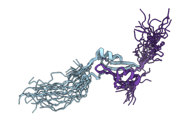
Isoreticular Co-Crystal Of Replication Initiator Protein Repe54 And Asymmetrical Expanded Duplex (31Mer) Containing The Cognate Repe54 Sequence, And Additional T-A Rich Sequence With 5' Terminal Phosphates. Two Copies Of Even-Skipped Homeodomain Loaded Post-Crystallization
Organism: Escherichia coli, Drosophila melanogaster
Method: X-RAY DIFFRACTION Resolution:3.02 Å Release Date: 2026-02-18 Classification: DNA BINDING PROTEIN/DNA Ligands: MG |
 |
Isoreticular Co-Crystal 1 With Asymmetrical Expanded Duplex (31Mer) Containing Insert Sequence Ctaattaggc And Loaded With Even-Skipped Homeodomain
Organism: Escherichia coli, Drosophila melanogaster
Method: X-RAY DIFFRACTION Resolution:3.07 Å Release Date: 2026-02-18 Classification: DNA BINDING PROTEIN Ligands: MG |
 |
Isoreticular Co-Crystal 1 With Asymmetrical Expanded Duplex (31Mer) Containing Insert Sequence Tgatgagcag And Loaded With Even-Skipped Homeodomain
Organism: Escherichia coli, Drosophila melanogaster
Method: X-RAY DIFFRACTION Resolution:3.16 Å Release Date: 2026-02-18 Classification: DNA BINDING PROTEIN Ligands: MG |
 |
Isoreticular Co-Crystal 1 With Asymmetrical Expanded Duplex (31Mer) Containing Insert Sequence Tgatgagcag And Loaded With Even-Skipped Homeodomain
Organism: Escherichia coli, Drosophila melanogaster
Method: X-RAY DIFFRACTION Resolution:2.96 Å Release Date: 2026-02-18 Classification: DNA BINDING PROTEIN Ligands: MG |
 |
Isoreticular Co-Crystal 1 With Asymmetrical Expanded Duplex (31Mer) Containing Insert Sequence Tgatgagcag And Loaded With Ultrabithorax Homeodomain
Organism: Drosophila melanogaster, Escherichia coli
Method: X-RAY DIFFRACTION Resolution:3.67 Å Release Date: 2026-02-18 Classification: DNA BINDING PROTEIN Ligands: MG |
 |
Isoreticular Co-Crystal 1 With Asymmetrical Expanded Duplex (31Mer) Containing Insert Sequence Tgatgagcag And Loaded With Engrailed Homeodomain Enhanced Green Fluorescent Protein Fusion
Organism: Escherichia coli, Drosophila melanogaster, Aequorea victoria
Method: X-RAY DIFFRACTION Resolution:3.75 Å Release Date: 2026-02-18 Classification: DNA BINDING PROTEIN/DNA Ligands: MG |
 |
Isoreticular Co-Crystal 1 With Asymmetrical Expanded Duplex (31Mer) Containing Insert Sequence Taattaggccg And Loaded With Ultrabithorax Homeodomain
Organism: Escherichia coli, Drosophila melanogaster
Method: X-RAY DIFFRACTION Resolution:3.39 Å Release Date: 2026-02-18 Classification: DNA BINDING PROTEIN Ligands: MG |
 |
Isoreticular Co-Crystal 1 With Asymmetrical Expanded Duplex (31Mer) Containing Insert Sequence Taattaggccg And Loaded With Even-Skipped Homeodomain
Organism: Escherichia coli, Drosophila melanogaster
Method: X-RAY DIFFRACTION Resolution:3.08 Å Release Date: 2026-02-18 Classification: DNA BINDING PROTEIN Ligands: MG |
 |
Isoreticular Co-Crystal 1 With Asymmetrical Expanded Duplex (31Mer) Containing Insert Sequence Taattaggccg And Loaded With Antennapedia Homeodomain
Organism: Escherichia coli, Drosophila melanogaster
Method: X-RAY DIFFRACTION Resolution:3.30 Å Release Date: 2026-02-18 Classification: DNA BINDING PROTEIN Ligands: MG |
 |
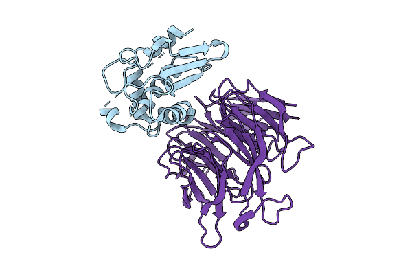
Structure Of The Proline-Rich Binding Domain Of Tesup-1 In Complex With Dzfc3H1 Peptide
Organism: Drosophila melanogaster
Method: X-RAY DIFFRACTION Resolution:1.90 Å Release Date: 2026-02-04 Classification: PROTEIN BINDING Ligands: TLA, ACT |
 |
Structure Of The Tesup-1 Proline-Rich Binding Domain In Complex With The Proline-Rich Region Of Pih1D1
Organism: Drosophila melanogaster
Method: X-RAY DIFFRACTION Resolution:2.10 Å Release Date: 2026-02-04 Classification: PROTEIN BINDING Ligands: GOL |
 |
Organism: Drosophila melanogaster
Method: SOLUTION NMR Release Date: 2026-01-28 Classification: METAL BINDING PROTEIN Ligands: ZN |
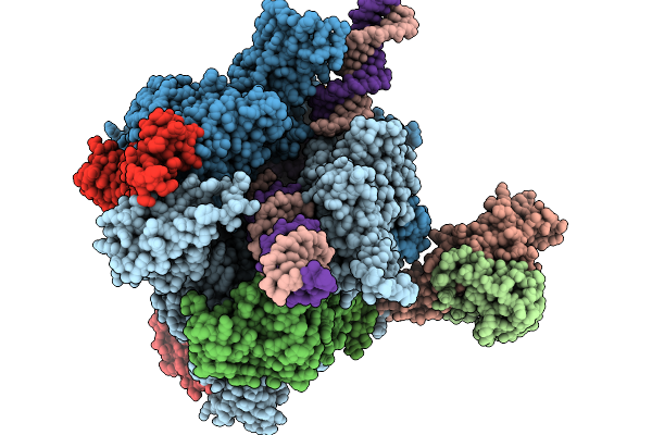 |
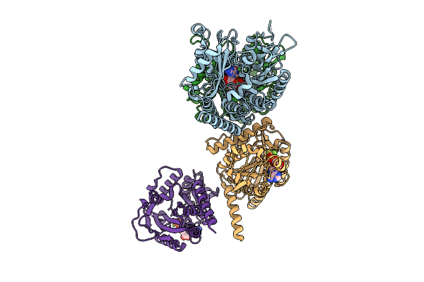
Structure Of A Native Drosophila Melanogaster Pol Ii Elongation Complex With A Well-Defined Rpb4/Rpb7 Stalk
Organism: Drosophila melanogaster
Method: ELECTRON MICROSCOPY Resolution:3.44 Å Release Date: 2026-01-07 Classification: TRANSCRIPTION Ligands: ZN |
 |
Organism: Drosophila melanogaster
Method: X-RAY DIFFRACTION Resolution:2.50 Å Release Date: 2025-12-24 Classification: METAL BINDING PROTEIN |
 |
Organism: Drosophila melanogaster
Method: ELECTRON MICROSCOPY Release Date: 2025-12-03 Classification: MEMBRANE PROTEIN Ligands: K |
 |
Organism: Drosophila melanogaster, Homo sapiens
Method: ELECTRON MICROSCOPY Release Date: 2025-10-29 Classification: LIGASE Ligands: AMP |
 |
Organism: Drosophila melanogaster, Homo sapiens
Method: ELECTRON MICROSCOPY Release Date: 2025-10-29 Classification: LIGASE Ligands: AMP |
 |
Organism: Drosophila melanogaster, Homo sapiens
Method: ELECTRON MICROSCOPY Release Date: 2025-10-29 Classification: LIGASE Ligands: AMP |
 |
Organism: Drosophila melanogaster, Homo sapiens
Method: ELECTRON MICROSCOPY Release Date: 2025-10-22 Classification: MEMBRANE PROTEIN Ligands: ABU |
 |
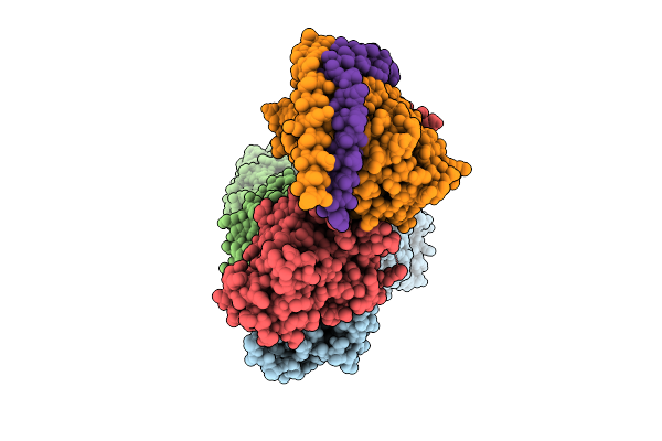
Organism: Drosophila melanogaster, Sus scrofa
Method: ELECTRON MICROSCOPY Release Date: 2025-10-15 Classification: CELL CYCLE Ligands: GTP, GDP, ADP, MG, ALF |