Search Count: 136
 |
Organism: Homo sapiens, Synthetic construct
Method: X-RAY DIFFRACTION Release Date: 2025-07-16 Classification: APOPTOSIS |
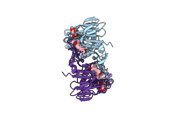 |
Organism: Severe acute respiratory syndrome coronavirus 2
Method: X-RAY DIFFRACTION Resolution:1.98 Å Release Date: 2025-04-09 Classification: HYDROLASE/HYDROLASE INHIBITOR Ligands: A1A0T, SIN |
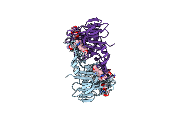 |
Organism: Severe acute respiratory syndrome coronavirus 2
Method: X-RAY DIFFRACTION Resolution:2.01 Å Release Date: 2025-04-09 Classification: HYDROLASE/HYDROLASE INHIBITOR Ligands: A1A0S, ACY, GOL |
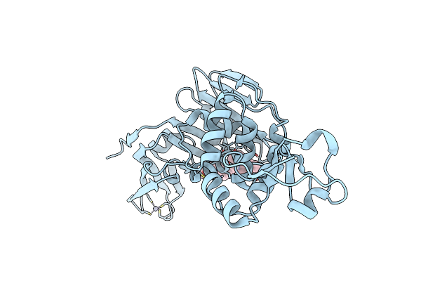 |
Organism: Severe acute respiratory syndrome coronavirus 2
Method: X-RAY DIFFRACTION Resolution:2.80 Å Release Date: 2025-04-09 Classification: HYDROLASE/HYDROLASE INHIBITOR Ligands: A1A0U, ZN, CL |
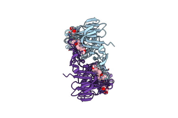 |
Organism: Severe acute respiratory syndrome coronavirus 2
Method: X-RAY DIFFRACTION Resolution:1.88 Å Release Date: 2025-04-09 Classification: HYDROLASE/HYDROLASE INHIBITOR Ligands: A1A0V, ACY, GOL |
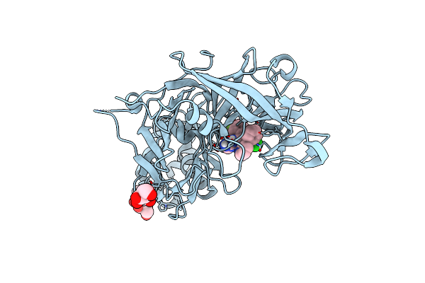 |
Organism: Plasmodium vivax sal-1
Method: X-RAY DIFFRACTION Release Date: 2024-08-28 Classification: HYDROLASE/Inhibitor Ligands: NAG, T0F, EDO |
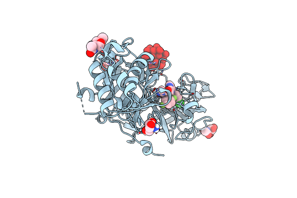 |
Organism: Plasmodium vivax sal-1
Method: X-RAY DIFFRACTION Release Date: 2024-08-28 Classification: HYDROLASE Ligands: SKC, TEW, NAG, TRS |
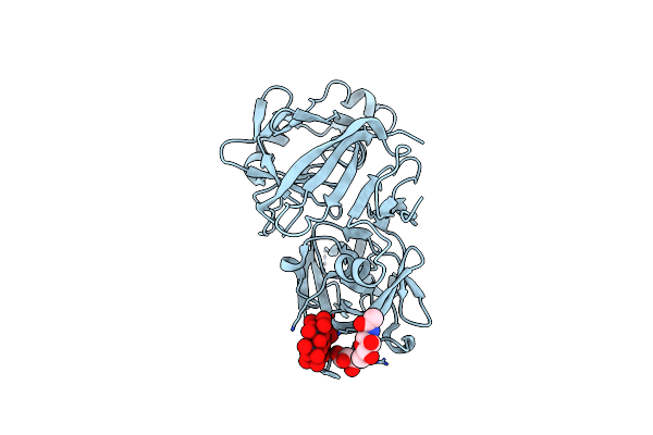 |
Organism: Plasmodium vivax sal-1
Method: X-RAY DIFFRACTION Release Date: 2024-08-28 Classification: HYDROLASE/Inhibitor Ligands: GOL, TEW, NAG |
 |
Organism: Homo sapiens
Method: X-RAY DIFFRACTION Resolution:2.40 Å Release Date: 2024-04-10 Classification: APOPTOSIS/INHIBITOR Ligands: SO4, CCN, PEG, 144, PGE, EDO, 1PE |
 |
Organism: Synthetic construct
Method: X-RAY DIFFRACTION Release Date: 2024-03-20 Classification: DE NOVO PROTEIN |
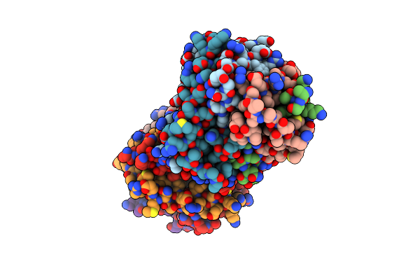 |
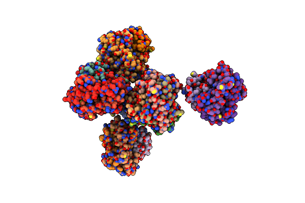
Crystal Structure Of Bax Core Domain Bh3-Groove Dimer - Tetrameric Fraction P21
Organism: Homo sapiens
Method: X-RAY DIFFRACTION Resolution:2.09 Å Release Date: 2023-12-27 Classification: APOPTOSIS Ligands: EDO |
 |
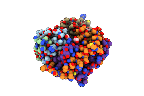
Crystal Structure Of Bax Core Domain Bh3-Groove Dimer - Tetrameric Fraction P31
Organism: Homo sapiens
Method: X-RAY DIFFRACTION Resolution:2.30 Å Release Date: 2023-12-27 Classification: APOPTOSIS Ligands: ZN, EDO, PEG |
 |
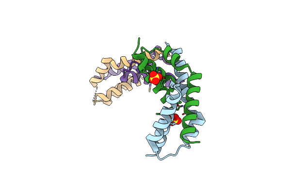
Crystal Structure Of Bax Core Domain Bh3-Groove Dimer - Hexameric Fraction With 2-Stearoyl Lysopc
Organism: Homo sapiens
Method: X-RAY DIFFRACTION Resolution:2.20 Å Release Date: 2023-12-27 Classification: APOPTOSIS Ligands: 1GP, EDO, OCT, D12, DD9 |
 |
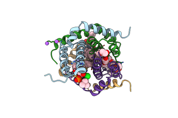
Crystal Structure Of Bax Core Domain Bh3-Groove Dimer - Hexameric Fraction With Dioctanoyl Phosphatidylserine
Organism: Homo sapiens
Method: X-RAY DIFFRACTION Resolution:2.40 Å Release Date: 2023-12-27 Classification: APOPTOSIS Ligands: SO4 |
 |
Organism: Homo sapiens
Method: X-RAY DIFFRACTION Resolution:2.09 Å Release Date: 2023-12-27 Classification: APOPTOSIS Ligands: K6G, ZN, NA, PEG, EDO, CL |
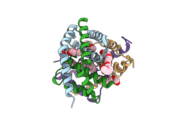 |
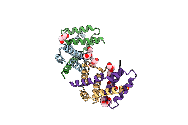
Organism: Homo sapiens
Method: X-RAY DIFFRACTION Resolution:2.40 Å Release Date: 2023-12-27 Classification: APOPTOSIS Ligands: PG0, PEG, N8E, P33, PG4 |
 |
Organism: Homo sapiens
Method: X-RAY DIFFRACTION Resolution:2.25 Å Release Date: 2023-12-27 Classification: APOPTOSIS Ligands: PEG, PGE, PG4, SO4 |
 |
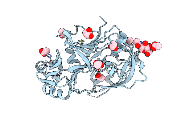
Crystal Structure Of Phosphorylated (T357/S358) Human Mlkl Pseudokinase Domain
Organism: Homo sapiens
Method: X-RAY DIFFRACTION Resolution:2.30 Å Release Date: 2023-11-08 Classification: TRANSFERASE |
 |
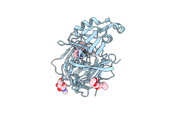
Organism: Plasmodium falciparum
Method: X-RAY DIFFRACTION Resolution:1.85 Å Release Date: 2022-05-04 Classification: HYDROLASE Ligands: NAG, ACT, GOL |
 |
Crystal Structure Of Plasmepsin X From Plasmodium Falciparum In Complex With Wm382
Organism: Plasmodium falciparum
Method: X-RAY DIFFRACTION Resolution:2.76 Å Release Date: 2022-05-04 Classification: HYDROLASE Ligands: EDO, NAG, PG4, I0L |

