
Search Count: 208
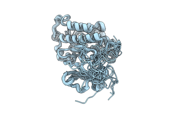 |
Organism: Homo sapiens
Method: SOLUTION NMR Release Date: 2026-01-21 Classification: SIGNALING PROTEIN |
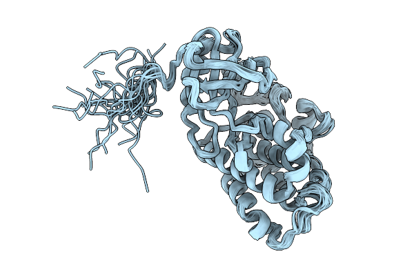 |
Organism: Homo sapiens
Method: SOLUTION NMR Release Date: 2026-01-21 Classification: SIGNALING PROTEIN |
 |
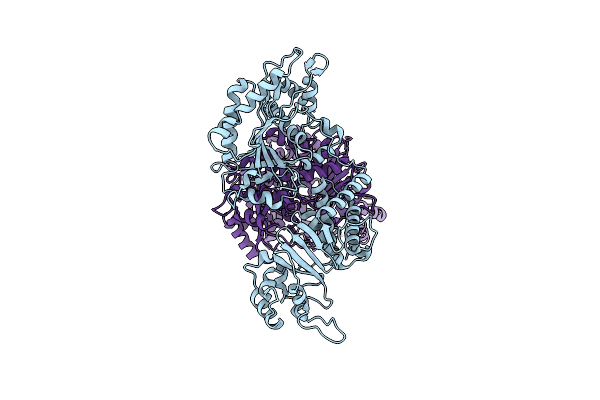
Organism: Escherichia coli
Method: ELECTRON MICROSCOPY Resolution:2.60 Å Release Date: 2025-09-17 Classification: RNA BINDING PROTEIN/DNA/RNA |
 |
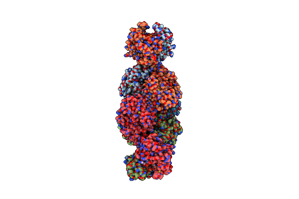
Organism: Homo sapiens
Method: X-RAY DIFFRACTION Resolution:2.30 Å Release Date: 2025-05-07 Classification: IMMUNE SYSTEM Ligands: CDP, A1L1P, ZN |
 |
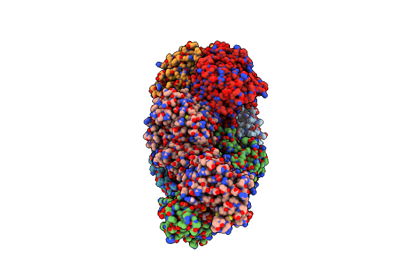
Cryo-Electron Microscopy (Cryo-Em) Structure Of The Hachiman Defense System From Escherichia Coli
Organism: Escherichia coli k-12
Method: ELECTRON MICROSCOPY Release Date: 2025-03-26 Classification: DNA BINDING PROTEIN |
 |
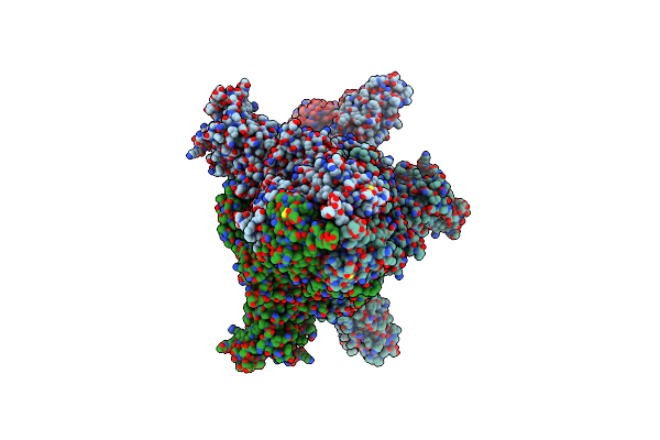
The Structure Of Type Iii Crispr-Associated Deaminase In Complex Ca6 And Atp, Fully Activated
Organism: Limisphaera ngatamarikiensis
Method: ELECTRON MICROSCOPY Resolution:3.40 Å Release Date: 2024-12-25 Classification: IMMUNE SYSTEM Ligands: ATP, ZN, MG |
 |
Organism: Limisphaera ngatamarikiensis
Method: ELECTRON MICROSCOPY Release Date: 2024-12-25 Classification: IMMUNE SYSTEM Ligands: ZN, LQJ |
 |
Organism: Escherichia coli
Method: ELECTRON MICROSCOPY Release Date: 2024-12-18 Classification: RNA BINDING PROTEIN/DNA/RNA Ligands: NCA, AR6 |
 |
Organism: Limisphaera ngatamarikiensis
Method: ELECTRON MICROSCOPY Release Date: 2024-12-11 Classification: IMMUNE SYSTEM Ligands: ZN, ATP |
 |
Organism: Limisphaera ngatamarikiensis
Method: ELECTRON MICROSCOPY Release Date: 2024-12-11 Classification: IMMUNE SYSTEM |
 |
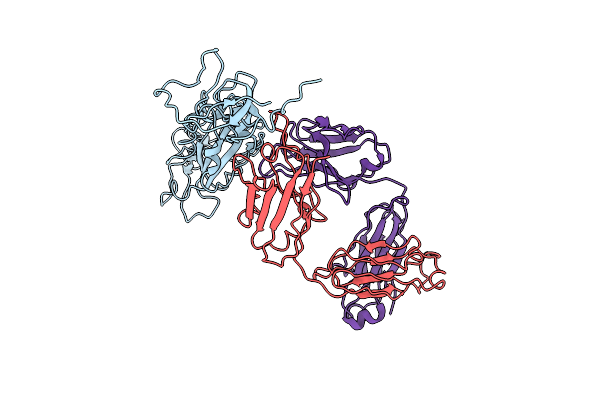
Crystal Structure Of Ttd-Phd Domain Of Uhrf1 In Complex With Mstella Peptide (Residues 85-119)
Organism: Homo sapiens, Mus musculus
Method: X-RAY DIFFRACTION Resolution:3.20 Å Release Date: 2024-11-06 Classification: GENE REGULATION/INHIBITOR Ligands: ZN |
 |
Crystal Structure Of Phd Domain Of Uhrf1 In Complex With Mstella Peptide (Residues 85-119)
Organism: Homo sapiens, Mus musculus
Method: X-RAY DIFFRACTION Resolution:1.60 Å Release Date: 2024-11-06 Classification: GENE REGULATION Ligands: SCN, GOL, ZN |
 |
Crystal Structure Of Ttd-Phd Domain Of Uhrf1 In Complex With Hstella Peptide (Residues 75-121)
Organism: Homo sapiens
Method: X-RAY DIFFRACTION Resolution:2.25 Å Release Date: 2024-11-06 Classification: GENE REGULATION Ligands: ZN, GOL, ACY |
 |
Crystal Structure Of Phd Domain Of Uhrf1 In Complex With Hstella Peptide (Residues 75-121)
Organism: Homo sapiens
Method: X-RAY DIFFRACTION Resolution:2.05 Å Release Date: 2024-11-06 Classification: GENE REGULATION Ligands: ZN |
 |
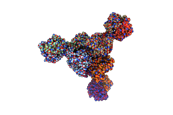
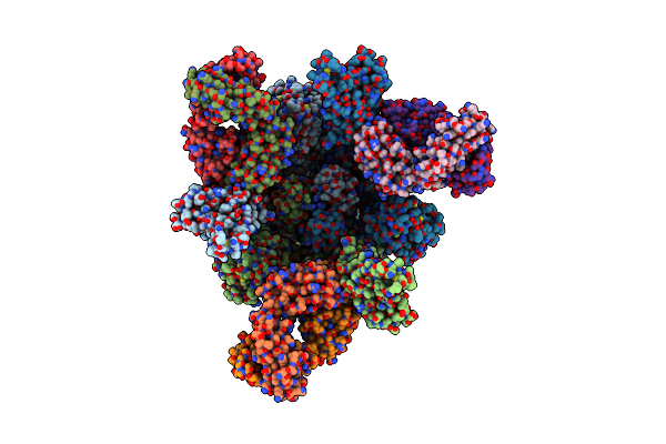
State 2 Of Sars-Cov-2 Xbb Variant Spike Protein Trimer Complexed With Antibody Pw5-5
Organism: Severe acute respiratory syndrome coronavirus 2, Homo sapiens
Method: ELECTRON MICROSCOPY Release Date: 2024-09-04 Classification: VIRAL PROTEIN/IMMUNE SYSTEM |
 |
Organism: Severe acute respiratory syndrome coronavirus 2, Homo sapiens
Method: ELECTRON MICROSCOPY Release Date: 2024-08-14 Classification: VIRAL PROTEIN/IMMUNE SYSTEM |
 |
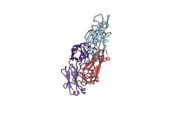
The Local Refined Map Of Sars-Cov-2 Xbb Variant Spike Protein Complexed With Antibody Pw5-535
Organism: Homo sapiens, Severe acute respiratory syndrome coronavirus 2
Method: ELECTRON MICROSCOPY Release Date: 2024-08-14 Classification: IMMUNE SYSTEM/VIRAL PROTEIN |
 |
Organism: Homo sapiens, Severe acute respiratory syndrome coronavirus 2
Method: ELECTRON MICROSCOPY Release Date: 2024-08-14 Classification: IMMUNE SYSTEM/VIRAL PROTEIN |
 |
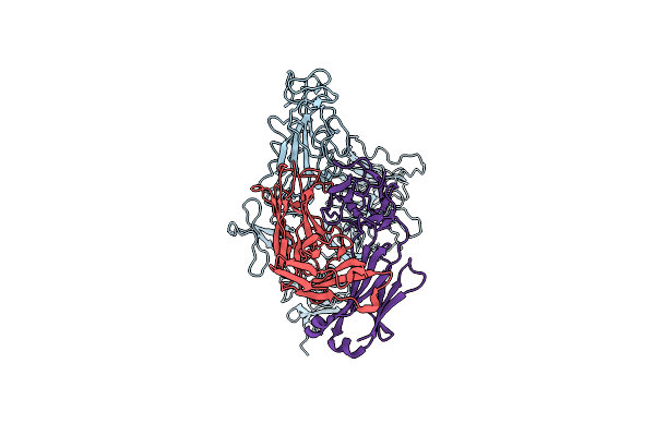
The Local Refined Map Of Sars-Cov Spike Protein Complexed With Antibody Pw5-5
Organism: Severe acute respiratory syndrome coronavirus 2, Homo sapiens
Method: ELECTRON MICROSCOPY Release Date: 2024-08-14 Classification: VIRAL PROTEIN/IMMUNE SYSTEM |
 |
Monomer State Of Sars-Cov-2 Xbb Variant Spike Protein Trimer Complexed With Antibody Pw5-5
Organism: Homo sapiens, Severe acute respiratory syndrome coronavirus 2
Method: ELECTRON MICROSCOPY Release Date: 2024-08-14 Classification: IMMUNE SYSTEM/VIRAL PROTEIN |