
Search Count: 96
 |
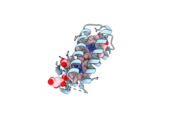
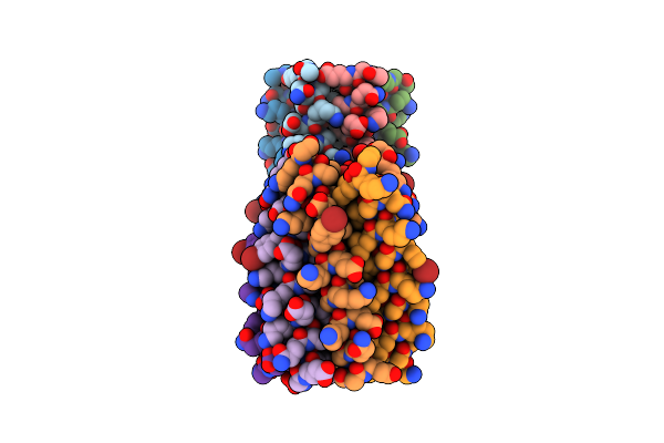
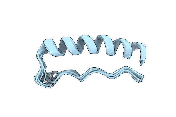
Solution Nmr Structure Of Lactamised Alpha-Synuclein 2-12 Peptide In 50% Tfe.
Organism: Homo sapiens
Method: SOLUTION NMR Release Date: 2024-07-10 Classification: LIPID BINDING PROTEIN |
 |
Organism: Homo sapiens
Method: X-RAY DIFFRACTION Resolution:1.78 Å Release Date: 2023-09-27 Classification: CELL CYCLE |
 |
Organism: Escherichia coli
Method: X-RAY DIFFRACTION Resolution:2.10 Å Release Date: 2023-08-09 Classification: ELECTRON TRANSPORT Ligands: HEM, LMR, CL |
 |
Organism: Homo sapiens
Method: SOLUTION NMR Release Date: 2023-08-09 Classification: LIPID BINDING PROTEIN |
 |
Organism: Micromonospora maris (strain dsm 45365 / jcm 31040 / nbrc 109089 / nrrl b-24793 / ab-18-032)
Method: X-RAY DIFFRACTION Resolution:2.01 Å Release Date: 2022-11-30 Classification: OXIDOREDUCTASE Ligands: HEM, PGE, GOL, PEG, P6G, P33, CL, MG |
 |
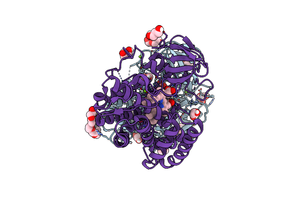
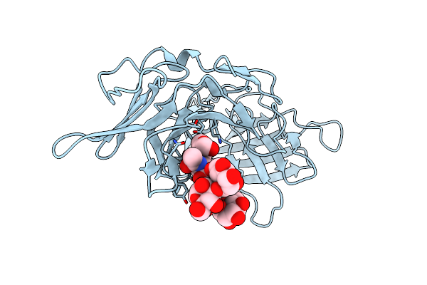
High Resolution Structure Of Alpha-1-Acid Glycoprotein Bound To Potent Anti-Tumour Compound Ucn-01
Organism: Homo sapiens
Method: X-RAY DIFFRACTION Resolution:1.82 Å Release Date: 2021-12-01 Classification: PROTEIN TRANSPORT Ligands: UCN |
 |
Organism: Synthetic construct
Method: X-RAY DIFFRACTION Resolution:1.91 Å Release Date: 2021-10-06 Classification: DE NOVO PROTEIN Ligands: HEM |
 |
Organism: Synthetic construct
Method: X-RAY DIFFRACTION Resolution:2.01 Å Release Date: 2021-02-03 Classification: DE NOVO PROTEIN |
 |
Organism: Homo sapiens
Method: X-RAY DIFFRACTION Resolution:1.50 Å Release Date: 2020-08-19 Classification: SUGAR BINDING PROTEIN |
 |
Organism: Homo sapiens
Method: X-RAY DIFFRACTION Resolution:2.56 Å Release Date: 2020-08-19 Classification: SUGAR BINDING PROTEIN Ligands: CL, NA |
 |
Organism: Homo sapiens
Method: X-RAY DIFFRACTION Resolution:3.47 Å Release Date: 2020-08-19 Classification: SUGAR BINDING PROTEIN Ligands: SO4, NAG |
 |
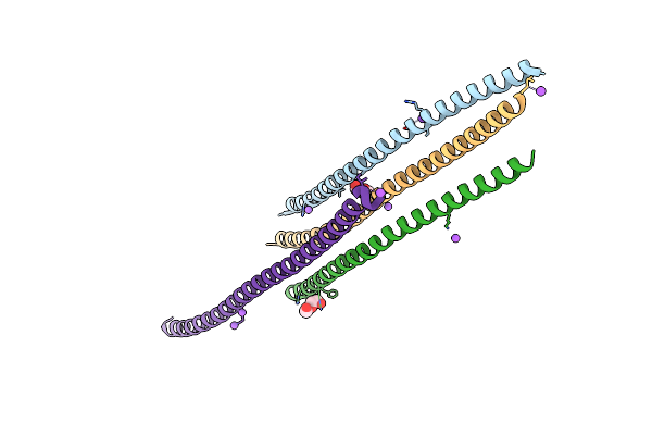
Nmr Structure Of Repeat Domain 13 Of The Fibrillar Adhesin Csha From Streptococcus Gordonii.
Organism: Streptococcus gordonii (strain challis / atcc 35105 / bcrc 15272 / ch1 / dl1 / v288)
Method: SOLUTION NMR Release Date: 2020-04-08 Classification: CELL ADHESION |
 |
Crystal Structure Of S. Aureus Fabi In Complex With Nadph And Kalimantacin A (Batumin)
Organism: Staphylococcus aureus
Method: X-RAY DIFFRACTION Resolution:2.45 Å Release Date: 2020-04-01 Classification: BIOSYNTHETIC PROTEIN Ligands: NDP, KAL |
 |
Crystal Structure Of S. Aureus Fabi In Complex With Nadph And Kalimantacin B
Organism: Staphylococcus aureus
Method: X-RAY DIFFRACTION Resolution:2.55 Å Release Date: 2020-04-01 Classification: BIOSYNTHETIC PROTEIN Ligands: NDP, N5H |
 |
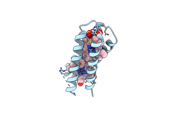
Solution Structure Of Macpd, A Acyl Carrier Protein, From Pseudomonas Fluorescens Involved In Mupirocin Biosynthesis.
Organism: Pseudomonas fluorescens
Method: SOLUTION NMR Release Date: 2020-02-19 Classification: BIOSYNTHETIC PROTEIN |
 |
Organism: Streptococcus mutans
Method: SOLUTION NMR Release Date: 2019-07-10 Classification: STRUCTURAL PROTEIN |
 |
Organism: Pseudomonas fluorescens
Method: X-RAY DIFFRACTION Resolution:1.45 Å Release Date: 2019-03-06 Classification: BIOSYNTHETIC PROTEIN |
 |
Solution Structure Of The Apo Doublet Acyl Carrier Protein From Prodigiosin Biosynthesis
Organism: serratia sp. atcc 39006
Method: SOLUTION NMR Release Date: 2018-12-12 Classification: BIOSYNTHETIC PROTEIN |
 |
Crystal Structure Of The N-Terminal Domain Of Burkholderia Pseudomallei Antitoxin Hicb
Organism: Burkholderia pseudomallei k96243
Method: X-RAY DIFFRACTION Resolution:1.56 Å Release Date: 2018-10-31 Classification: ANTITOXIN |
 |
Organism: Burkholderia pseudomallei
Method: X-RAY DIFFRACTION Resolution:1.85 Å Release Date: 2018-10-31 Classification: ANTITOXIN Ligands: CL, GOL |