
Search Count: 44
All
Selected
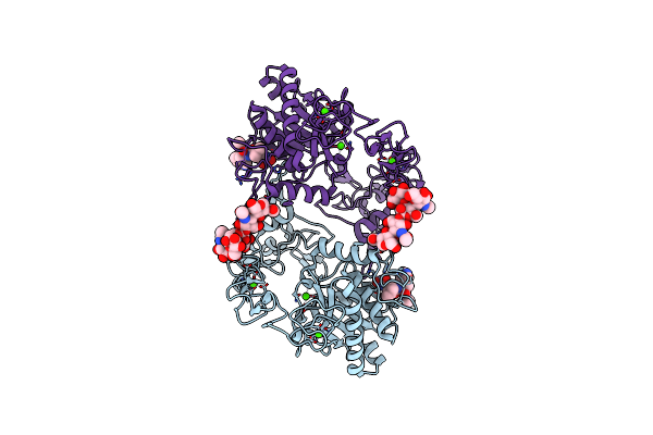 |
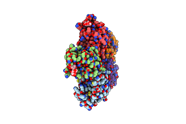
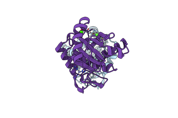
Organism: Crotalus atrox
Method: X-RAY DIFFRACTION Resolution:2.15 Å Release Date: 2007-07-10 Classification: APOPTOSIS, TOXIN Ligands: ZN, CA, GM6 |
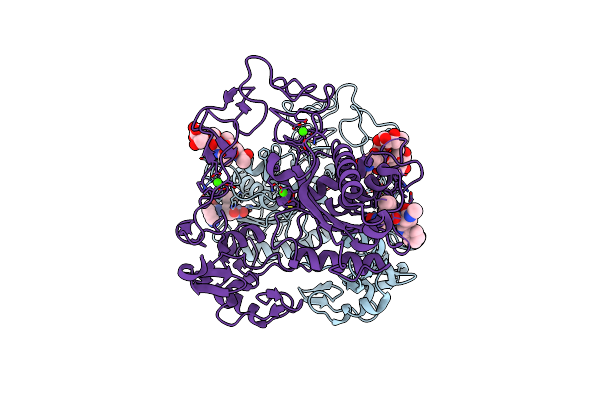 |
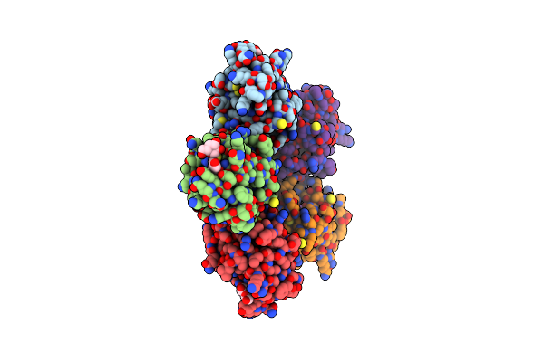
Organism: Crotalus atrox
Method: X-RAY DIFFRACTION Resolution:2.50 Å Release Date: 2007-07-10 Classification: APOPTOSIS, TOXIN Ligands: CA, ZN, GM6 |
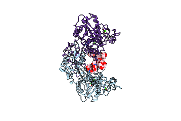 |
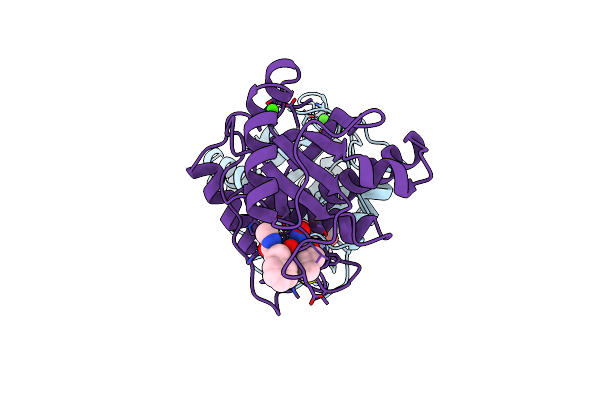
Organism: Crotalus atrox
Method: X-RAY DIFFRACTION Resolution:2.70 Å Release Date: 2007-07-10 Classification: APOPTOSIS, TOXIN Ligands: ZN, CA |
 |
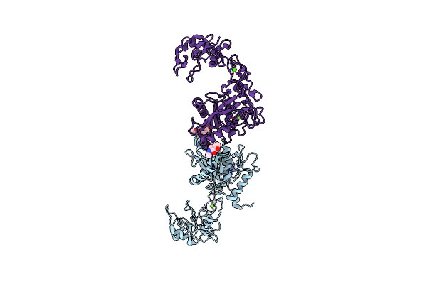
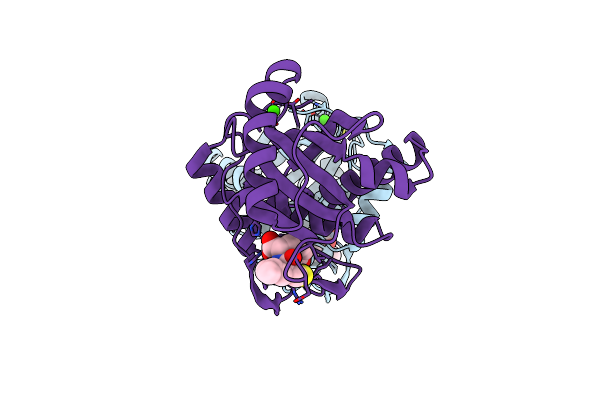
Crystal Structure Of Vascular Apoptosis-Inducing Protein-1(Orthorhombic Crystal Form)
Organism: Crotalus atrox
Method: X-RAY DIFFRACTION Resolution:2.50 Å Release Date: 2006-06-20 Classification: TOXIN Ligands: ZN, CA, 3CO |
 |
Crystal Structure Of Vascular Apoptosis-Inducing Protein-1(Inhibitor-Bound Form)
Organism: Crotalus atrox
Method: X-RAY DIFFRACTION Resolution:2.95 Å Release Date: 2006-06-20 Classification: TOXIN Ligands: ZN, CA, 3CO, GM6 |
 |
Crystal Structure Of Vascular Apoptosis-Inducing Protein-1(Tetragonal Crystal Form)
Organism: Crotalus atrox
Method: X-RAY DIFFRACTION Resolution:2.50 Å Release Date: 2006-06-20 Classification: TOXIN Ligands: ZN, CA, NAG |
 |
Organism: Crotalus atrox
Method: X-RAY DIFFRACTION Resolution:2.20 Å Release Date: 2003-07-01 Classification: SUGAR BINDING PROTEIN Ligands: CA, NA, CL |
 |
X-Ray Crystal Structure Of Rattlesnake Venom Complexed With Thiodigalactoside
Organism: Crotalus atrox
Method: X-RAY DIFFRACTION Resolution:2.30 Å Release Date: 2003-07-01 Classification: SUGAR BINDING PROTEIN Ligands: CA, NA, GAL |
 |
Organism: Crotalus atrox
Method: X-RAY DIFFRACTION Resolution:2.00 Å Release Date: 1997-02-12 Classification: HYDROLASE Ligands: ZN, CA, BAT |
 |
Structural Interaction Of Natural And Synthetic Inhibitors With The Venom Metalloproteinase, Atrolysin C (Form-D)
Organism: Crotalus atrox
Method: X-RAY DIFFRACTION Resolution:1.80 Å Release Date: 1995-10-15 Classification: HYDROLASE/HYDROLASE INHIBITOR Ligands: ZN, CA, 0QI |
 |
Structural Interaction Of Natural And Synthetic Inhibitors With The Venom Metalloproteinase, Atrolysin C (Ht-D)
Organism: Crotalus atrox
Method: X-RAY DIFFRACTION Resolution:2.10 Å Release Date: 1995-09-15 Classification: METALLOPROTEASE Ligands: ZN, CA |
 |
The Refined Crystal Structure Of Dimeric Phospholipase A2 At 2.5 Angstroms. Access To A Shielded Catalytic Center
Organism: Crotalus atrox
Method: X-RAY DIFFRACTION Resolution:2.50 Å Release Date: 1986-05-07 Classification: HYDROLASE |
 |
NA
|
 |
NA
|
 |
NA
|
 |
NA
|
 |
NA
|
 |
NA
|
 |
NA
|
 |
NA
|