
Search Count: 19
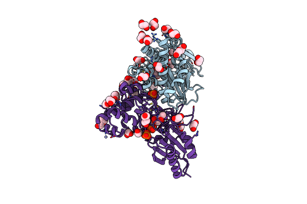 |
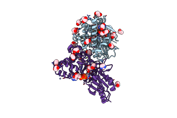
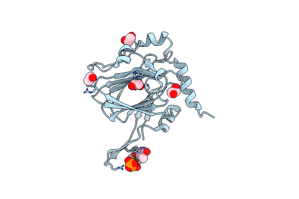
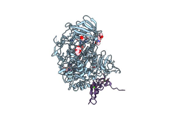
N-Acetylglucosamine Kinase From Plesiomonas Shigelloides Compexed With Alpha-N-Acetylglucosamine-6-Phosphate
Organism: Saccharomyces cerevisiae (strain atcc 204508 / s288c), Plesiomonas shigelloides 302-73
Method: X-RAY DIFFRACTION Resolution:1.75 Å Release Date: 2022-08-10 Classification: SUGAR BINDING PROTEIN Ligands: EDO, PEG, 4QY, ZN, K, PO4, CL, TRS |
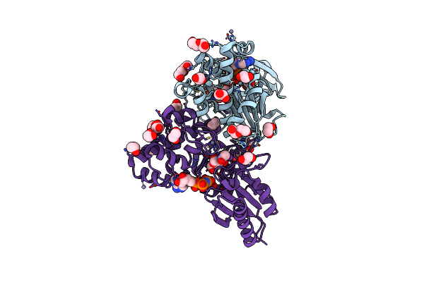 |
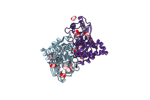
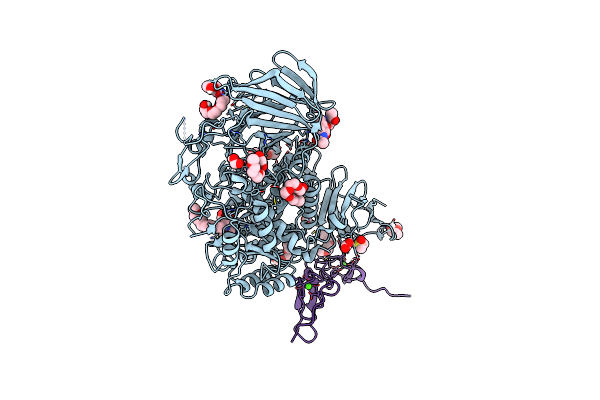
N-Acetylglucosamine Kinase From Plesiomonas Shigelloides Compexed With Alpha-N-Acetylglucosamine And Amp-Pnp Inhibitor
Organism: Saccharomyces cerevisiae (strain atcc 204508 / s288c), Plesiomonas shigelloides 302-73
Method: X-RAY DIFFRACTION Resolution:2.11 Å Release Date: 2022-08-10 Classification: SUGAR BINDING PROTEIN Ligands: ZN, ANP, PEG, PGE, NDG, EDO, K |
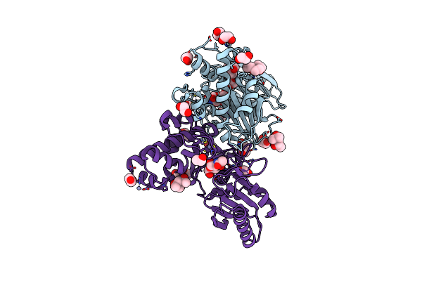 |
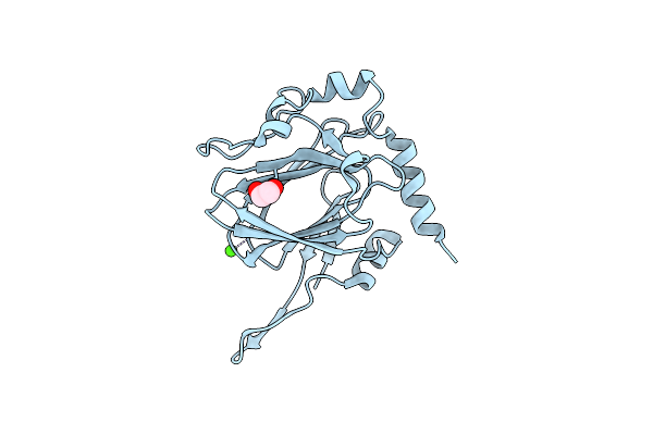
N-Acetylglucosamine Kinase From Plesiomonas Shigelloides Compexed With Alpha-N-Acetylglucosamine
Organism: Saccharomyces cerevisiae (strain atcc 204508 / s288c), Plesiomonas shigelloides 302-73
Method: X-RAY DIFFRACTION Resolution:1.94 Å Release Date: 2022-08-10 Classification: SUGAR BINDING PROTEIN Ligands: MPD, EDO, PGE, NDG, ZN, K, CL, PEG |
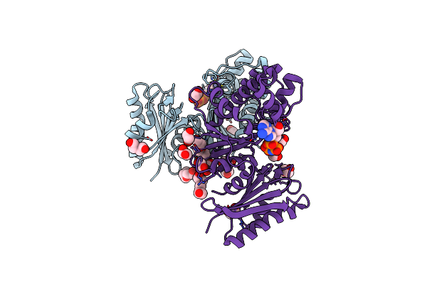 |
Structure Of N-Acetylglucosamine Kinase From Plesiomonas Shigelloides In Complex With Amp-Pnp In The Absence Of N-Acetylglucoseamine Substrate
Organism: Saccharomyces cerevisiae (strain atcc 204508 / s288c), Plesiomonas shigelloides 302-73
Method: X-RAY DIFFRACTION Resolution:2.20 Å Release Date: 2022-08-10 Classification: SUGAR BINDING PROTEIN Ligands: PEG, ANP, ZN, PGE, EDO |
 |
N-Acetylglucosamine Kinase From Plesiomonas Shigelloides Compexed With Alpha-N-Acetylglucosamine And Adp
Organism: Saccharomyces cerevisiae (strain atcc 204508 / s288c), Plesiomonas shigelloides 302-73
Method: X-RAY DIFFRACTION Resolution:1.57 Å Release Date: 2022-08-03 Classification: SUGAR BINDING PROTEIN Ligands: ADP, GOL, IMD, NDG, ZN, EDO, PEG, K, IPA |
 |
Native Structure Of N-Acetylglucosamine Kinase From Plesiomonas Shigelloides
Organism: Saccharomyces cerevisiae (strain atcc 204508 / s288c), Plesiomonas shigelloides 302-73
Method: X-RAY DIFFRACTION Resolution:1.70 Å Release Date: 2022-07-27 Classification: SUGAR BINDING PROTEIN Ligands: PEG, TRS, ZN, PGE, EDO, CL |
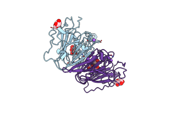 |
Organism: Coxiella burnetii (strain rsa 493 / nine mile phase i)
Method: X-RAY DIFFRACTION Resolution:1.43 Å Release Date: 2022-04-20 Classification: SUGAR BINDING PROTEIN Ligands: XYP, XYS, NA, FLC |
 |
Organism: Coxiella burnetii (strain rsa 493 / nine mile phase i)
Method: X-RAY DIFFRACTION Resolution:1.87 Å Release Date: 2022-04-20 Classification: SUGAR BINDING PROTEIN Ligands: TYD, CIT, EDO |
 |
Organism: Streptomyces griseus
Method: X-RAY DIFFRACTION Resolution:1.90 Å Release Date: 2022-04-20 Classification: SUGAR BINDING PROTEIN Ligands: EDO, TYD, CL |
 |
Organism: Streptomyces griseus
Method: X-RAY DIFFRACTION Resolution:1.33 Å Release Date: 2022-04-20 Classification: SUGAR BINDING PROTEIN Ligands: EDO, CA |
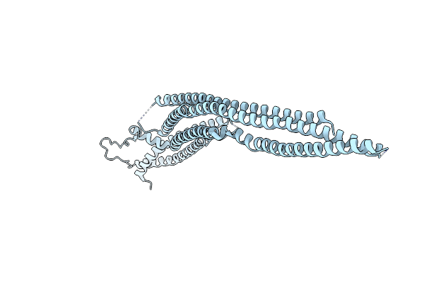 |
Organism: Homo sapiens
Method: X-RAY DIFFRACTION Resolution:2.70 Å Release Date: 2017-03-08 Classification: SIGNALING PROTEIN |
 |
Organism: Mus musculus
Method: X-RAY DIFFRACTION Resolution:1.74 Å Release Date: 2016-07-27 Classification: HYDROLASE Ligands: PG4, P6G, EDO, FMT, CA |
 |
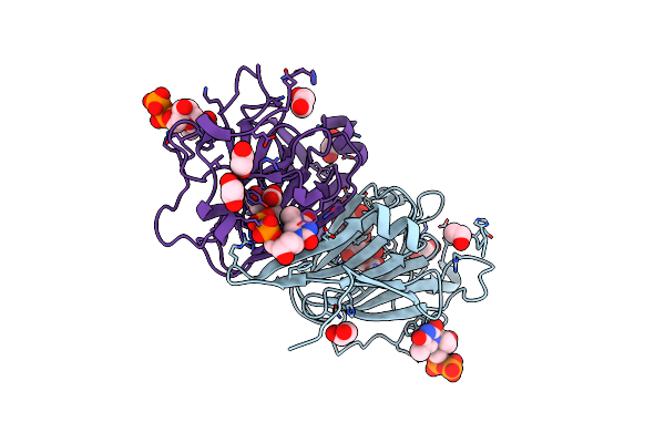
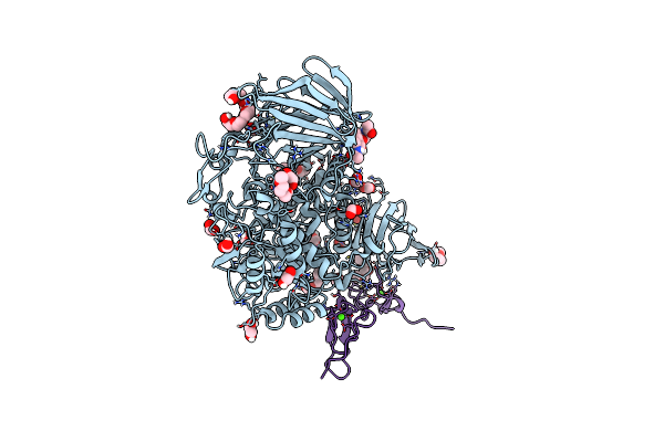
Complex Of Murine Endoplasmic Reticulum Alpha-Glucosidase Ii With D-Glucose
Organism: Mus musculus
Method: X-RAY DIFFRACTION Resolution:2.37 Å Release Date: 2016-07-27 Classification: HYDROLASE Ligands: NAG, EDO, FMT, PG4, P6G, OXM, GLC, CA, TAR |
 |
Murine Endoplasmic Reticulum Alpha-Glucosidase Ii With Bound Substrate Analogue
Organism: Mus musculus
Method: X-RAY DIFFRACTION Resolution:2.29 Å Release Date: 2016-07-27 Classification: HYDROLASE Ligands: ACT, EDO, FMT, DGO, CA |
 |
Murine Endoplasmic Reticulum Alpha-Glucosidase Ii With Bound Covalent Intermediate
Organism: Mus musculus
Method: X-RAY DIFFRACTION Resolution:2.40 Å Release Date: 2016-07-27 Classification: HYDROLASE Ligands: EDO, FMT, 5GF, P6G, OXM, TAR, CA |
 |
Organism: Mus musculus
Method: X-RAY DIFFRACTION Resolution:1.81 Å Release Date: 2016-07-27 Classification: HYDROLASE Ligands: PG4, P6G, EDO, FMT, CTS, CA |
 |
Organism: Mus musculus
Method: X-RAY DIFFRACTION Resolution:1.92 Å Release Date: 2016-07-27 Classification: HYDROLASE Ligands: PG4, P6G, EDO, FMT, NOJ, CA |
 |
Murine Endoplasmic Reticulum Alpha-Glucosidase Ii With N-Butyl-1-Deoxynojirimycin
Organism: Mus musculus
Method: X-RAY DIFFRACTION Resolution:2.38 Å Release Date: 2016-07-27 Classification: HYDROLASE Ligands: FMT, ACT, NBV, P6G, CA |
 |
Murine Endoplasmic Reticulum Alpha-Glucosidase Ii With N-9'-Methoxynonyl-1-Deoxynojirimycin.
Organism: Mus musculus
Method: X-RAY DIFFRACTION Release Date: 2016-07-27 Classification: HYDROLASE Ligands: 6A9, FMT, P6G, DMS, PG4, CA |