
Search Count: 86
 |
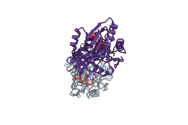
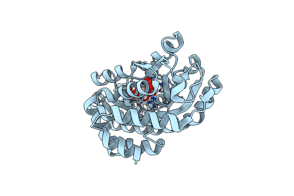
Organism: Aquifex aeolicus vf5
Method: X-RAY DIFFRACTION Resolution:1.60 Å Release Date: 2022-02-02 Classification: TRANSFERASE Ligands: E8Q, CO3, SAH, SAM |
 |
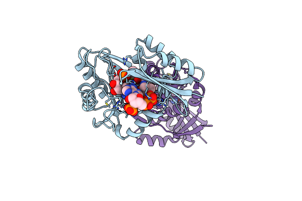
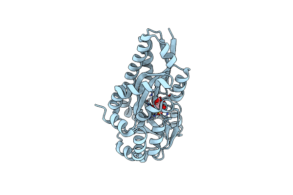
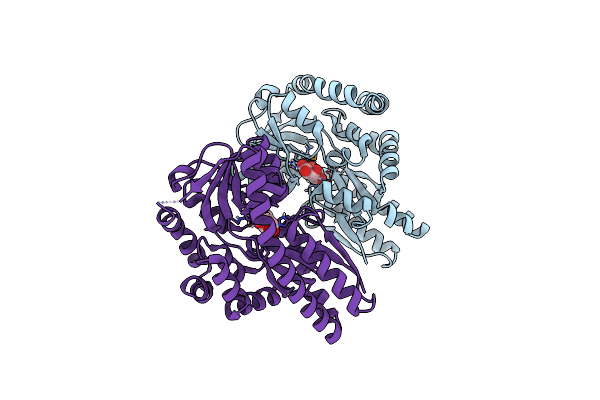
Organism: Helicobacter pylori
Method: X-RAY DIFFRACTION Resolution:1.70 Å Release Date: 2021-12-01 Classification: FLAVOPROTEIN Ligands: SF4, FMN, CL |
 |
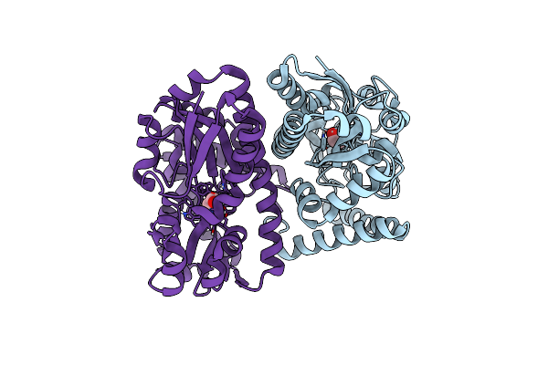
Crystal Structure Of Dehydrogenase/Isomerase Fabx From Helicobacter Pylori In Complex With Holo-Acp
Organism: Helicobacter pylori
Method: X-RAY DIFFRACTION Resolution:2.80 Å Release Date: 2021-12-01 Classification: FLAVOPROTEIN Ligands: FMN, SF4, PN7 |
 |
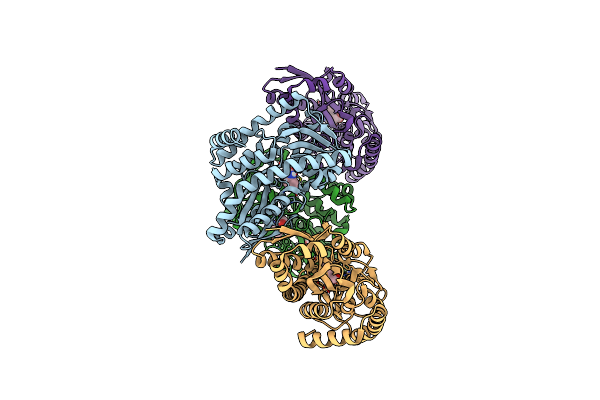
Crystal Structure Of Dehydrogenase/Isomerase Fabx From Helicobacter Pylori In Complex With Octanoyl-Acp
Organism: Helicobacter pylori
Method: X-RAY DIFFRACTION Resolution:2.31 Å Release Date: 2021-12-01 Classification: FLAVOPROTEIN Ligands: FMN, SF4, 66S |
 |
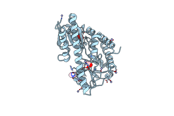
Crystal Structure Of Biog From Haemophilus Influenzae At 1.26 Angstroms Resolution
Organism: Haemophilus influenzae (strain atcc 51907 / dsm 11121 / kw20 / rd)
Method: X-RAY DIFFRACTION Resolution:1.26 Å Release Date: 2016-12-07 Classification: HYDROLASE |
 |
Crystal Structure Of Semet-Biog From Haemophilus Influenzae At 1.49 Angstroms Resolution
Organism: Haemophilus influenzae rd kw20
Method: X-RAY DIFFRACTION Resolution:1.49 Å Release Date: 2016-12-07 Classification: HYDROLASE Ligands: GOL, IPA |
 |
Organism: Aquifex aeolicus
Method: X-RAY DIFFRACTION Resolution:2.50 Å Release Date: 2016-12-07 Classification: LIGASE |
 |
Organism: Aquifex aeolicus
Method: X-RAY DIFFRACTION Resolution:2.46 Å Release Date: 2016-12-07 Classification: LIGASE Ligands: PML |
 |
Organism: Aquifex aeolicus
Method: X-RAY DIFFRACTION Resolution:2.55 Å Release Date: 2016-12-07 Classification: LIGASE Ligands: APC, MG, PML |
 |
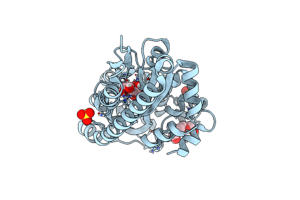
Organism: Aquifex aeolicus
Method: X-RAY DIFFRACTION Resolution:2.25 Å Release Date: 2016-12-07 Classification: LIGASE Ligands: AMP, COA |
 |
Crystal Structure Of A Trap Periplasmic Solute Binding Protein From Chromohalobacter Salexigens Dsm 3043 (Csal_0678), Target Efi-501078, With Bound Ethanolamine
Organism: Chromohalobacter salexigens
Method: X-RAY DIFFRACTION Resolution:1.40 Å Release Date: 2014-09-03 Classification: TRANSPORT PROTEIN Ligands: CL, ETA |
 |
Crystal Structure Of A Trap Periplasmic Solute Binding Protein From Desulfovibrio Alaskensis G20 (Dde_0634, Target Efi-510120) With Bound 3-Indole Acetic Acid
Organism: Desulfovibrio desulfuricans
Method: X-RAY DIFFRACTION Resolution:2.25 Å Release Date: 2014-06-18 Classification: SOLUTE-BINDING PROTEIN Ligands: IAC, EDO |
 |
Crystal Structure Of A Trap Periplasmic Solute Binding Protein From Desulfovibrio Alaskensis G20 (Dde_0634, Target Efi-510120) With Bound Indole Pyruvate
Organism: Desulfovibrio desulfuricans
Method: X-RAY DIFFRACTION Resolution:1.80 Å Release Date: 2014-05-28 Classification: TRANSPORT PROTEIN Ligands: 3IO, EDO |
 |
Crystal Structure Of A Trap Periplasmic Solute Binding Protein From Rosenbacter Denitrificans Och 114 (Rd1_1052, Target Efi-510238) With Bound D-Mannonate
Organism: Roseobacter denitrificans
Method: X-RAY DIFFRACTION Resolution:1.30 Å Release Date: 2014-05-21 Classification: SOLUTE-BINDING PROTEIN Ligands: CS2 |
 |
Crystal Structure Of A Trap Periplasmic Solute Binding Protein From Polaromonas Sp Js666 (Bpro_0088, Target Efi-510167) Bound To D-Erythronate
Organism: Polaromonas sp. js666
Method: X-RAY DIFFRACTION Resolution:1.70 Å Release Date: 2014-05-14 Classification: SOLUTE-BINDING PROTEIN Ligands: EAX, MLA, FMT |
 |
Crystal Structure Of A Trap Periplasmic Solute Binding Protein From Polaromonas Sp Js666 (Bpro_1871, Target Efi-510164) Bound To D-Erythronate
Organism: Polaromonas sp. js666
Method: X-RAY DIFFRACTION Resolution:1.80 Å Release Date: 2014-05-14 Classification: SOLUTE-BINDING PROTEIN Ligands: EAX |
 |
Crystal Structure Of A Trap Periplasmic Solute Binding Protein From Rhodobacter Sphaeroides (Rsph17029_3620, Target Efi-510199), Apo Open Structure
Organism: Rhodobacter sphaeroides
Method: X-RAY DIFFRACTION Resolution:1.35 Å Release Date: 2014-05-14 Classification: SOLUTE-BINDING PROTEIN Ligands: IMD, EDO, ACT, ZN |
 |
Crystal Structure Of A Trap Periplasmic Solute Binding Protein From Colwellia Psychrerythraea (Cps_0129, Target Efi-510097) With Bound Calcium And Pyruvate
Organism: Colwellia psychrerythraea
Method: X-RAY DIFFRACTION Resolution:1.90 Å Release Date: 2014-05-14 Classification: SOLUTE-BINDING PROTEIN Ligands: CL, PYR, CA |
 |
Crystal Structure Of A Trap Periplasmic Solute Binding Protein From Roseobacter Denitrificans (Rd1_0742, Target Efi-510239) With Bound 3-Deoxy-D-Manno-Oct-2-Ulosonic Acid (Kdo)
Organism: Roseobacter denitrificans
Method: X-RAY DIFFRACTION Resolution:1.75 Å Release Date: 2014-05-14 Classification: TRANSPORT PROTEIN Ligands: KDO, PGE, SO4 |
 |
Crystal Structure Of A Trap Periplasmic Solute Binding Protein From Sulfitobacter Sp. Nas-14.1 (Target Efi-510299) With Bound Beta-D-Galacturonate
Organism: Sulfitobacter sp. nas-14.1
Method: X-RAY DIFFRACTION Resolution:1.50 Å Release Date: 2014-05-14 Classification: TRANSPORT PROTEIN Ligands: CL, GTR |