
Search Count: 23
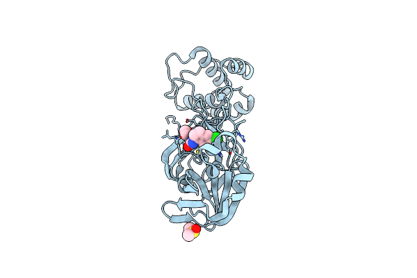 |
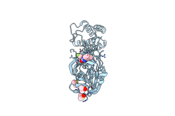
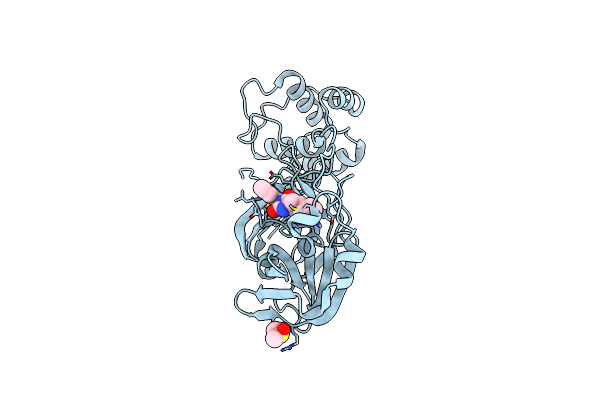
Crystal Structure Of Sars-Cov-2 Main Protease (Nsp5) In Complex With Compound 3
Organism: Severe acute respiratory syndrome coronavirus 2
Method: X-RAY DIFFRACTION Resolution:1.82 Å Release Date: 2022-02-23 Classification: VIRAL PROTEIN Ligands: DMS, RY5 |
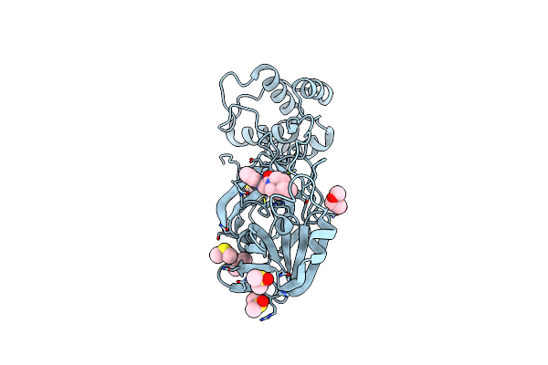 |
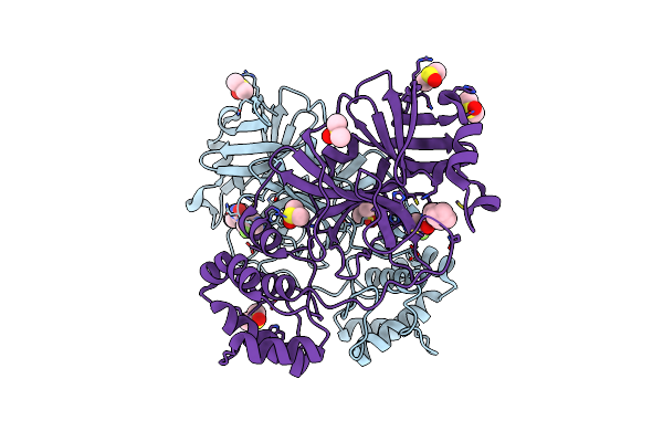
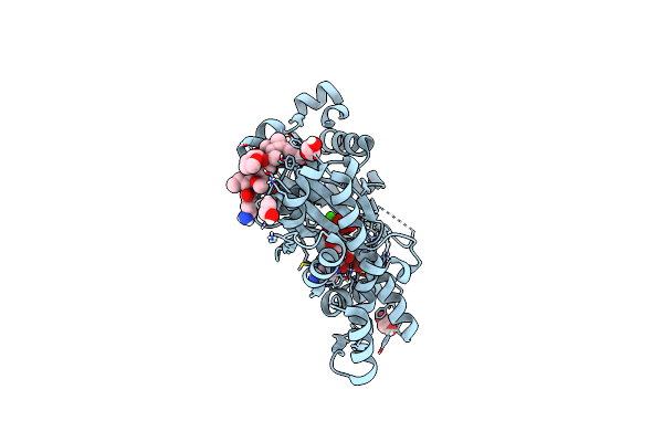
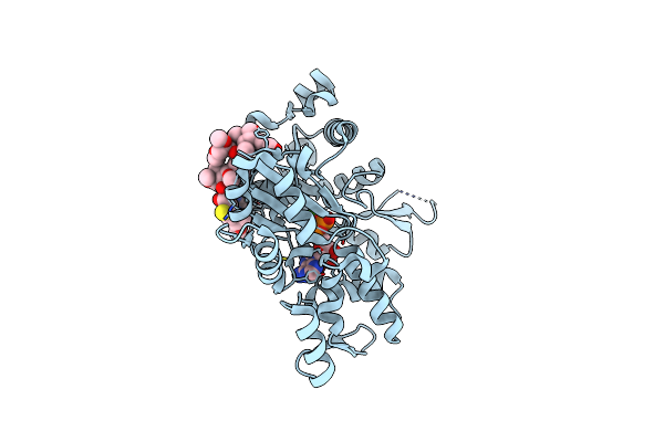
Crystal Structure Of Sars-Cov-2 Main Protease (Nsp5) In Complex With Compound 5
Organism: Severe acute respiratory syndrome coronavirus 2
Method: X-RAY DIFFRACTION Resolution:1.55 Å Release Date: 2022-02-23 Classification: VIRAL PROTEIN Ligands: SQ2, PEG, DMS |
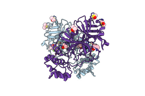 |
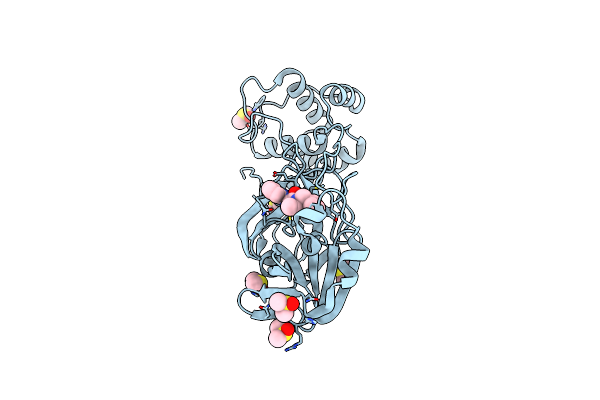
Crystal Structure Of Sars-Cov-2 Main Protease (Nsp5) In Complex With Compound 1
Organism: Severe acute respiratory syndrome coronavirus 2
Method: X-RAY DIFFRACTION Resolution:1.55 Å Release Date: 2022-02-23 Classification: VIRAL PROTEIN Ligands: SQ5, DMS |
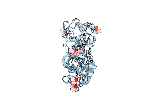 |
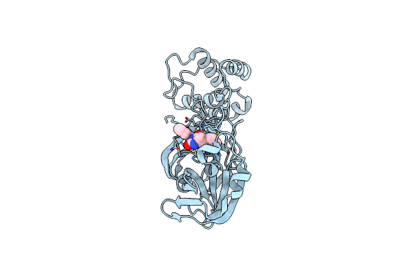
Crystal Structure Of Sars-Cov-2 Main Protease (Nsp5) In Complex With Compound 6
Organism: Severe acute respiratory syndrome coronavirus 2
Method: X-RAY DIFFRACTION Resolution:1.65 Å Release Date: 2022-02-23 Classification: VIRAL PROTEIN Ligands: DMS, SYH |
 |
Crystal Structure Of Sars-Cov-2 Main Protease (Nsp5) In Complex With Compound 8
Organism: Severe acute respiratory syndrome coronavirus 2
Method: X-RAY DIFFRACTION Resolution:1.60 Å Release Date: 2022-02-23 Classification: VIRAL PROTEIN Ligands: DMS, T0W |
 |
Crystal Structure Of Sars-Cov-2 Main Protease (Nsp5) In Complex With Compound 13
Organism: Severe acute respiratory syndrome coronavirus 2
Method: X-RAY DIFFRACTION Resolution:1.47 Å Release Date: 2022-02-23 Classification: VIRAL PROTEIN Ligands: DMS, TU8 |
 |
Crystal Structure Of Sars-Cov-2 Main Protease (Nsp5) In Complex With Compound 21
Organism: Severe acute respiratory syndrome coronavirus 2
Method: X-RAY DIFFRACTION Resolution:1.63 Å Release Date: 2022-02-23 Classification: VIRAL PROTEIN Ligands: DMS, U7W |
 |
Crystal Structure Of Sars-Cov-2 Main Protease (Nsp5) In Complex With Compound 15
Organism: Severe acute respiratory syndrome coronavirus 2
Method: X-RAY DIFFRACTION Resolution:1.64 Å Release Date: 2022-02-23 Classification: VIRAL PROTEIN Ligands: U9H, DMS |
 |
Crystal Structure Of Sars-Cov-2 Main Protease (Nsp5) In Complex With Compound 17
Organism: Severe acute respiratory syndrome coronavirus 2
Method: X-RAY DIFFRACTION Resolution:2.23 Å Release Date: 2022-02-23 Classification: VIRAL PROTEIN Ligands: V18 |
 |
Crystal Structure Of Sars-Cov-2 Main Protease (Nsp5) In Complex With Compound 18
Organism: Severe acute respiratory syndrome coronavirus 2
Method: X-RAY DIFFRACTION Resolution:2.00 Å Release Date: 2022-02-23 Classification: VIRAL PROTEIN Ligands: DMS, V1B |
 |
Organism: Oryctolagus cuniculus
Method: X-RAY DIFFRACTION Resolution:1.70 Å Release Date: 2020-10-21 Classification: CONTRACTILE PROTEIN Ligands: CA, ATP, LAB, EDO, TFJ |
 |
Organism: Plasmodium falciparum
Method: X-RAY DIFFRACTION Resolution:2.17 Å Release Date: 2019-09-25 Classification: PROTEIN BINDING |
 |
Organism: Plasmodium falciparum, Homo sapiens
Method: X-RAY DIFFRACTION Resolution:3.67 Å Release Date: 2019-09-25 Classification: CELL ADHESION Ligands: NAG |
 |
Organism: Oryctolagus cuniculus
Method: X-RAY DIFFRACTION Resolution:2.20 Å Release Date: 2018-11-21 Classification: STRUCTURAL PROTEIN Ligands: JQV, CA, ADP |
 |
Organism: Gallus gallus
Method: X-RAY DIFFRACTION Resolution:3.20 Å Release Date: 2017-08-23 Classification: CELL ADHESION Ligands: NAG |
 |
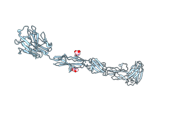
Crystal Structure Of The Chicken Mdga1 Ectodomain In Complex With The Human Neuroligin 1 (Nl1(-A-B)) Cholinesterase Domain.
Organism: Homo sapiens, Gallus gallus
Method: X-RAY DIFFRACTION Resolution:3.30 Å Release Date: 2017-08-23 Classification: CELL ADHESION Ligands: NAG |
 |
Crystal Structure Of The Human Neuroligin 1 Cholinesterase Domain Containing Spliced Sequence B (Ssb) (Nl1(-A+B))
Organism: Homo sapiens
Method: X-RAY DIFFRACTION Resolution:2.55 Å Release Date: 2017-08-23 Classification: CELL ADHESION Ligands: NAG, PGE |
 |
Crystal Structure Of Chicken Receptor Protein Tyrosine Phosphatase Sigma In Complex With Trkc
Organism: Gallus gallus
Method: X-RAY DIFFRACTION Resolution:2.50 Å Release Date: 2014-11-12 Classification: SIGNALING PROTEIN Ligands: NAG, SO4 |
 |
Crystal Structure Of Chicken Receptor Protein Tyrosine Phosphatase Sigma In Complex With Trkc
Organism: Gallus gallus
Method: X-RAY DIFFRACTION Resolution:3.05 Å Release Date: 2014-11-12 Classification: SIGNALING PROTEIN Ligands: NAG |
 |
Crystal Structure Of The Six N-Terminal Domains Of Human Receptor Protein Tyrosine Phosphatase Sigma
Organism: Homo sapiens
Method: X-RAY DIFFRACTION Resolution:3.15 Å Release Date: 2014-11-12 Classification: HYDROLASE Ligands: NAG |