
Search Count: 25
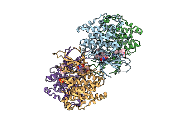 |
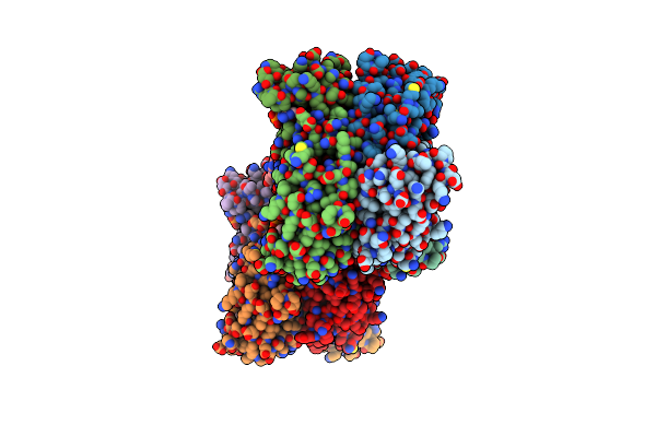
Toxoplasma Gondii Gsk3B Bound To Ly2090314 And Disulphide Bonded Through The C223 Residue
Organism: Toxoplasma gondii rh
Method: X-RAY DIFFRACTION Resolution:2.90 Å Release Date: 2025-10-22 Classification: TRANSFERASE Ligands: A1IYF, PO4 |
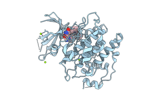 |
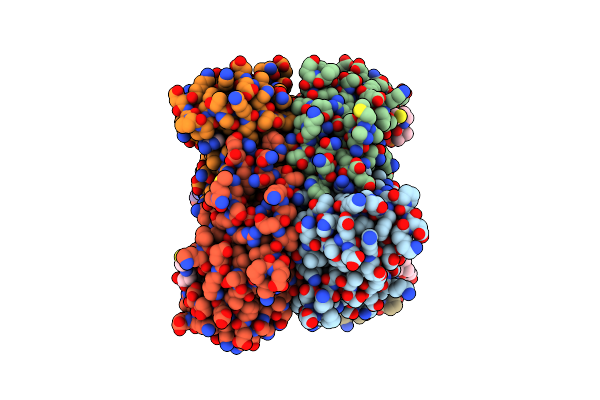
Organism: Toxoplasma gondii rh
Method: X-RAY DIFFRACTION Resolution:2.10 Å Release Date: 2025-10-15 Classification: TRANSFERASE Ligands: A1IYF, MG |
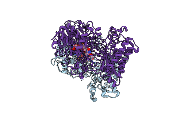 |
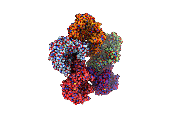
Knockout Of Gmc-Oxidoreductase Genes Reveals That Functional Redundancy Preserves Mimivirus Essential Functions
Organism: Mimivirus reunion
Method: ELECTRON MICROSCOPY Release Date: 2024-04-17 Classification: STRUCTURAL PROTEIN Ligands: FAD |
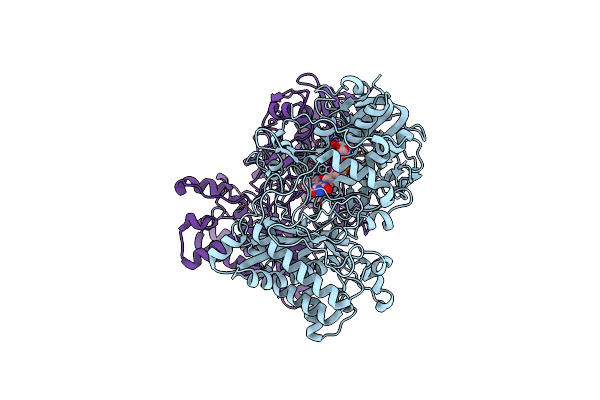 |
Knockout Of Gmc-Oxidoreductase Genes Reveals That Functional Redundancy Preserves Mimivirus Essential Functions
Organism: Mimivirus reunion
Method: ELECTRON MICROSCOPY Release Date: 2024-04-17 Classification: STRUCTURAL PROTEIN Ligands: FAD |
 |
Organism: Mus musculus
Method: X-RAY DIFFRACTION Resolution:1.96 Å Release Date: 2023-08-09 Classification: TRANSFERASE Ligands: SCA |
 |
Organism: Mus musculus
Method: X-RAY DIFFRACTION Resolution:2.20 Å Release Date: 2023-08-09 Classification: TRANSFERASE Ligands: ADP |
 |
Organism: Mus musculus
Method: X-RAY DIFFRACTION Resolution:2.60 Å Release Date: 2023-08-09 Classification: TRANSFERASE Ligands: COA |
 |
Organism: Homo sapiens
Method: ELECTRON MICROSCOPY Release Date: 2022-12-07 Classification: TRANSFERASE Ligands: NAG, UDP |
 |
Organism: Acanthamoeba polyphaga mimivirus
Method: ELECTRON MICROSCOPY Release Date: 2022-08-10 Classification: VIRAL PROTEIN Ligands: FAD |
 |
Organism: Acanthamoeba polyphaga mimivirus
Method: ELECTRON MICROSCOPY Release Date: 2022-08-10 Classification: VIRAL PROTEIN Ligands: FAD |
 |
Organism: Acanthamoeba polyphaga mimivirus
Method: ELECTRON MICROSCOPY Release Date: 2022-08-10 Classification: VIRAL PROTEIN Ligands: FAD |
 |
Organism: Acanthamoeba polyphaga mimivirus
Method: ELECTRON MICROSCOPY Release Date: 2022-08-10 Classification: VIRAL PROTEIN Ligands: FAD |
 |
Organism: Toxoplasma gondii
Method: X-RAY DIFFRACTION Resolution:2.31 Å Release Date: 2022-08-03 Classification: SPLICING Ligands: A9I, GOL, PEG, SCN |
 |
Organism: Toxoplasma gondii (strain atcc 50611 / me49)
Method: X-RAY DIFFRACTION Resolution:1.23 Å Release Date: 2021-07-21 Classification: RNA BINDING PROTEIN Ligands: IPA |
 |
Organism: Toxoplasma gondii (strain atcc 50611 / me49)
Method: X-RAY DIFFRACTION Resolution:1.35 Å Release Date: 2021-07-21 Classification: RNA BINDING PROTEIN Ligands: PG4, OYK |
 |
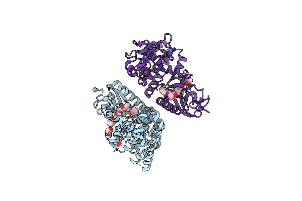
Crystal Structure Of The Toxoplasma Cpsf4 Yth-Domain In Complex With A 7 Mer M6A-Modified Rna
Organism: Toxoplasma gondii (strain atcc 50611 / me49), Synthetic construct
Method: X-RAY DIFFRACTION Resolution:1.38 Å Release Date: 2021-07-21 Classification: RNA BINDING PROTEIN Ligands: TOE, 6MD |
 |
Organism: Francisella tularensis subsp. novicida
Method: X-RAY DIFFRACTION Resolution:3.40 Å Release Date: 2021-01-27 Classification: LYASE |
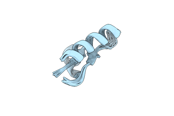 |
The Unusual Structure Of Ruminococcin C1 Antimicrobial Peptide Confers Activity Against Clinical Pathogens
Organism: [ruminococcus] gnavus e1
Method: SOLUTION NMR Release Date: 2020-08-12 Classification: ANTIMICROBIAL PROTEIN |
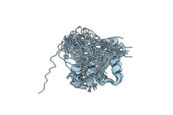 |
Organism: Bacillus subtilis
Method: SOLUTION NMR Release Date: 2020-01-08 Classification: MEMBRANE PROTEIN |
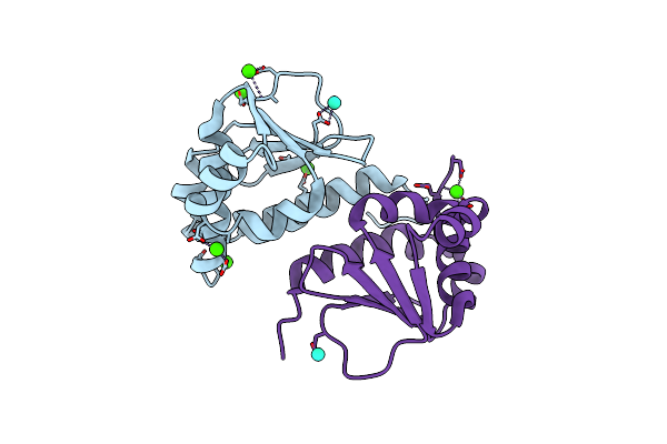 |
Organism: Escherichia coli (strain k12)
Method: X-RAY DIFFRACTION Resolution:2.12 Å Release Date: 2019-02-13 Classification: LIPID BINDING PROTEIN Ligands: TB, CA |