
Search Count: 5
 |
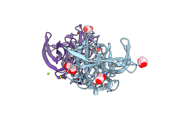
Two-Phospholipid-Bound Crystal Structure Of The Substrate-Binding Protein Ttg2D From Pseudomonas Aeruginosa
Organism: Pseudomonas aeruginosa pao1
Method: X-RAY DIFFRACTION Resolution:2.53 Å Release Date: 2019-10-23 Classification: LIPID TRANSPORT Ligands: GOT, H3T, GOL, SO4 |
 |
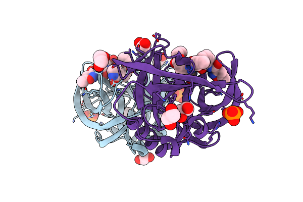
Crystal Structure Of C5321: A Protective Antigen Present In Uropathogenic Escherichia Coli Strains Displaying An Slr Fold
Organism: Escherichia coli
Method: X-RAY DIFFRACTION Resolution:1.74 Å Release Date: 2013-07-24 Classification: UNKNOWN FUNCTION Ligands: MG, CL, EDO |
 |
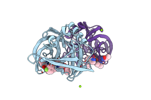
Organism: Human enterovirus b
Method: X-RAY DIFFRACTION Resolution:1.90 Å Release Date: 2011-09-07 Classification: HYDROLASE Ligands: GOL, MG |
 |
Organism: Human enterovirus b
Method: X-RAY DIFFRACTION Resolution:1.32 Å Release Date: 2011-09-07 Classification: HYDROLASE/HYDROLASE INHIBITOR Ligands: XNV, EDO, PI, NH4 |
 |
Organism: Human enterovirus b
Method: X-RAY DIFFRACTION Resolution:1.50 Å Release Date: 2011-09-07 Classification: HYDROLASE/HYDROLASE INHIBITOR Ligands: AG7, MG, CL |