
Search Count: 20
 |
Organism: Homo sapiens, Severe acute respiratory syndrome coronavirus 2
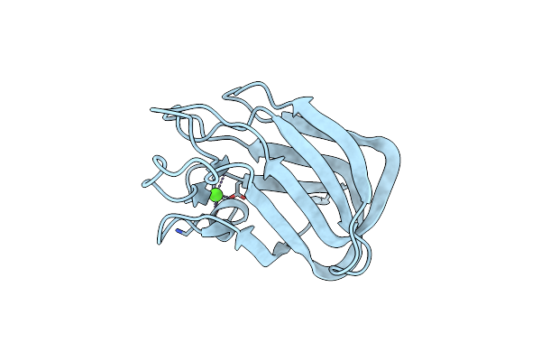
Method: X-RAY DIFFRACTION Resolution:1.67 Å Release Date: 2024-10-23 Classification: VIRAL PROTEIN Ligands: EDO, NAG, NI, TRS |
 |
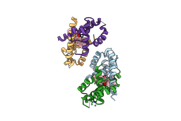
Vir-7229 Fab Fragment Bound The Sars-Cov-2 Ba.2.86 Spike Trimer (Local Refinement Of The Ba 2.86 Rbd/Vir-7229 Vhvl)
Organism: Homo sapiens
Method: ELECTRON MICROSCOPY Release Date: 2024-10-16 Classification: VIRAL PROTEIN/IMMUNE SYSTEM Ligands: NAG |
 |
Organism: Homo sapiens, Severe acute respiratory syndrome coronavirus 2
Method: X-RAY DIFFRACTION Resolution:1.90 Å Release Date: 2024-10-16 Classification: VIRAL PROTEIN Ligands: EDO, NI, CL, TRS, NAG |
 |
Organism: Homo sapiens, Severe acute respiratory syndrome coronavirus 2
Method: X-RAY DIFFRACTION Resolution:2.41 Å Release Date: 2024-10-16 Classification: VIRAL PROTEIN Ligands: K, EDO, ACY, EPE, NAG |
 |
Organism: Homo sapiens, Severe acute respiratory syndrome coronavirus 2
Method: ELECTRON MICROSCOPY Release Date: 2024-10-16 Classification: VIRAL PROTEIN/IMMUNE SYSTEM Ligands: NAG |
 |
Sars-Cov-2 Xbb.1 Spike Rbd Bound To The Human Ace2 Ectodomain And The S309 Neutralizing Antibody Fab Fragment
Organism: Homo sapiens, Severe acute respiratory syndrome coronavirus
Method: ELECTRON MICROSCOPY Release Date: 2023-10-04 Classification: VIRAL PROTEIN/IMMUNE SYSTEM Ligands: NAG |
 |
Sars-Cov-2 Bq.1.1 Spike Rbd Bound To The Human Ace2 Ectodomain And The S309 Neutralizing Antibody Fab Fragment
Organism: Homo sapiens, Severe acute respiratory syndrome coronavirus 2
Method: ELECTRON MICROSCOPY Release Date: 2023-10-04 Classification: VIRAL PROTEIN/IMMUNE SYSTEM Ligands: NAG, ZN |
 |
Sars-Cov-2 Bn.1 Spike Rbd Bound To The Human Ace2 Ectodomain And The S309 Neutralizing Antibody Fab Fragment
Organism: Homo sapiens, Severe acute respiratory syndrome coronavirus
Method: ELECTRON MICROSCOPY Release Date: 2023-10-04 Classification: VIRAL PROTEIN Ligands: NAG |
 |
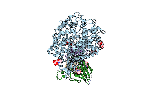
Organism: Mus musculus
Method: ELECTRON MICROSCOPY Release Date: 2023-09-20 Classification: HYDROLASE Ligands: PO4 |
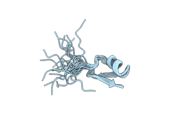 |
Organism: Myxococcus xanthus
Method: SOLUTION NMR Release Date: 2023-04-19 Classification: UNKNOWN FUNCTION |
 |
Crystal Structure Of The Multidrug Binding Transcriptional Regulator Lmrr In Complex Squaraine Dye
Organism: Lactococcus lactis subsp. lactis
Method: X-RAY DIFFRACTION Resolution:2.60 Å Release Date: 2022-06-01 Classification: FLUORESCENT PROTEIN Ligands: NI, 8TF |
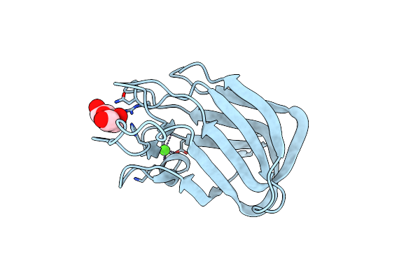 |
Structure Of Cbm Bt3015C From Bacteroides Thetaiotaomicron In Complex With Galactose
Organism: Bacteroides thetaiotaomicron (strain atcc 29148 / dsm 2079 / nctc 10582 / e50 / vpi-5482)
Method: X-RAY DIFFRACTION Resolution:1.18 Å Release Date: 2022-03-02 Classification: SUGAR BINDING PROTEIN Ligands: GAL, CA |
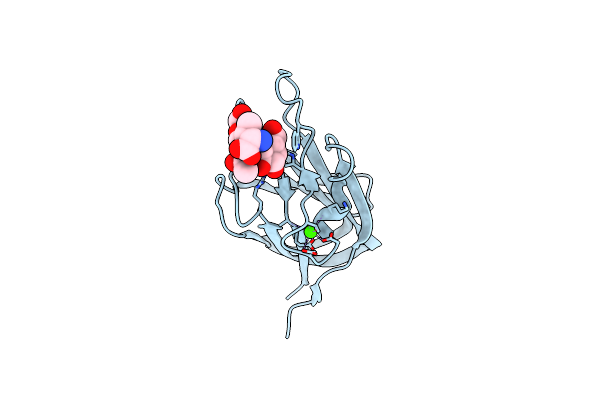 |
Structure Of Cbm Bt3015C From Bacteroides Thetaiotaomicron In Complex With O-Galnac Core 1-Thr
Organism: Bacteroides thetaiotaomicron vpi-5482
Method: X-RAY DIFFRACTION Resolution:1.79 Å Release Date: 2022-03-02 Classification: SUGAR BINDING PROTEIN Ligands: THR, CA |
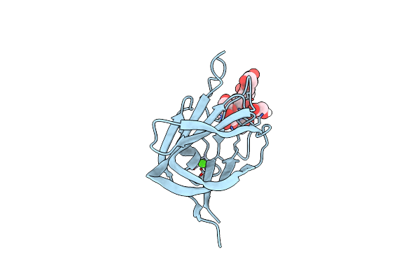 |
Structure Of Cbm Bt3015C From Bacteroides Thetaiotaomicron In Complex With O-Galnac Core 2-Thr
Organism: Bacteroides thetaiotaomicron (strain atcc 29148 / dsm 2079 / nctc 10582 / e50 / vpi-5482)
Method: X-RAY DIFFRACTION Resolution:1.76 Å Release Date: 2022-03-02 Classification: SUGAR BINDING PROTEIN Ligands: EDO, THR, CA |
 |
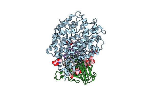
Organism: Bacteroides thetaiotaomicron (strain atcc 29148 / dsm 2079 / nctc 10582 / e50 / vpi-5482)
Method: X-RAY DIFFRACTION Resolution:1.06 Å Release Date: 2022-03-02 Classification: SUGAR BINDING PROTEIN Ligands: CA |
 |
Organism: Bacteroides thetaiotaomicron (strain atcc 29148 / dsm 2079 / nctc 10582 / e50 / vpi-5482)
Method: X-RAY DIFFRACTION Resolution:1.76 Å Release Date: 2022-03-02 Classification: SUGAR BINDING PROTEIN Ligands: PEG, EDO |
 |
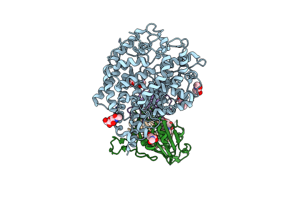
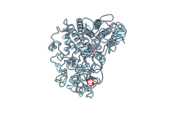
Organism: Homo sapiens
Method: X-RAY DIFFRACTION Resolution:2.80 Å Release Date: 2021-10-27 Classification: HYDROLASE Ligands: 85T, DTP, MG, GTP |
 |
Crystal Structure Of Cruzain In Complex With The Non-Covalent Inhibitor Nequimed176
Organism: Trypanosoma cruzi
Method: X-RAY DIFFRACTION Resolution:2.62 Å Release Date: 2013-09-18 Classification: HYDROLASE/HYDROLASE INHIBITOR Ligands: 1RV |
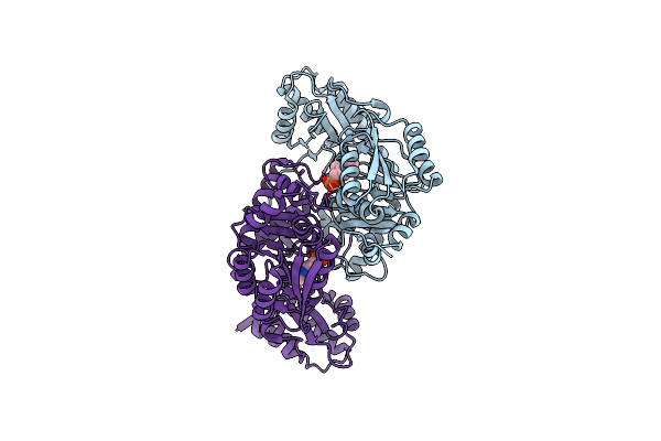 |
Crystal Structure Of Ureidoglycine-Glyoxylate Aminotransferase (Pucg) From Bacillus Subtilis
Organism: Bacillus subtilis
Method: X-RAY DIFFRACTION Resolution:2.06 Å Release Date: 2010-09-15 Classification: TRANSFERASE Ligands: PLP |
 |