
Search Count: 24
 |
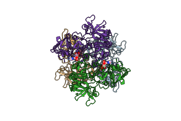
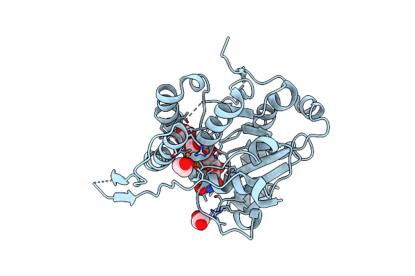
Organism: Escherichia coli
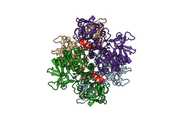
Method: ELECTRON MICROSCOPY Release Date: 2020-02-05 Classification: TRANSFERASE Ligands: FBP |
 |
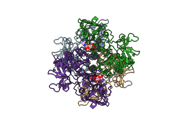
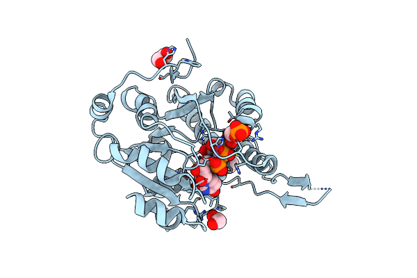
Organism: Escherichia coli
Method: ELECTRON MICROSCOPY Release Date: 2020-02-05 Classification: TRANSFERASE Ligands: AMP |
 |
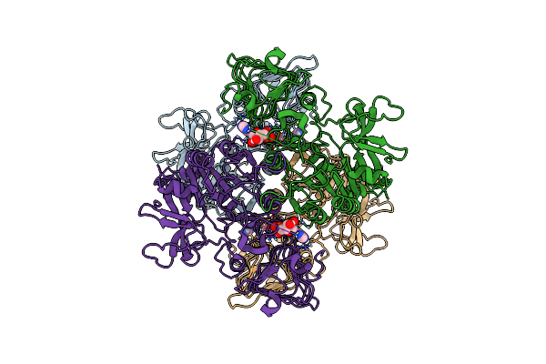
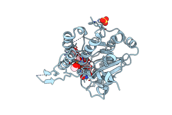
Organism: Escherichia coli
Method: ELECTRON MICROSCOPY Release Date: 2020-02-05 Classification: TRANSFERASE Ligands: FBP |
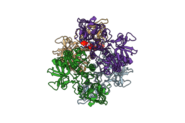 |
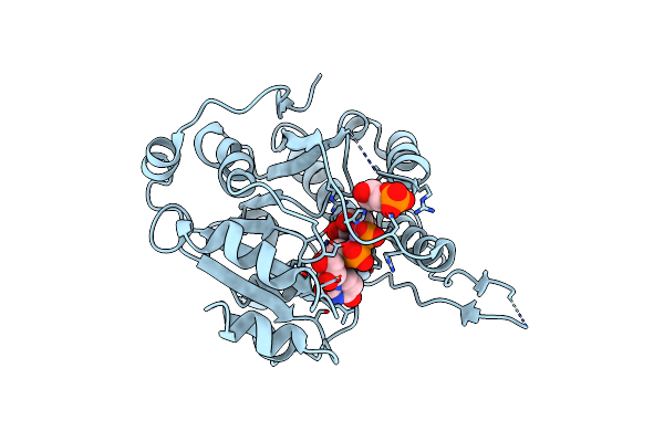
Organism: Escherichia coli
Method: ELECTRON MICROSCOPY Release Date: 2020-02-05 Classification: TRANSFERASE Ligands: FBP |
 |
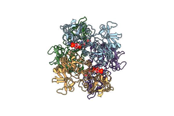
Organism: Escherichia coli
Method: ELECTRON MICROSCOPY Release Date: 2020-02-05 Classification: TRANSFERASE Ligands: AMP |
 |
Organism: Escherichia coli
Method: ELECTRON MICROSCOPY Release Date: 2020-02-05 Classification: TRANSFERASE Ligands: AMP |
 |
Crystal Structure Of Glucosyl-3-Phosphoglycerate Synthase From Mycobacterium Tuberculosis In Complex With Phosphoglyceric Acid (Pga) - Gpgs*Pga
Organism: Mycobacterium tuberculosis h37rv
Method: X-RAY DIFFRACTION Resolution:2.82 Å Release Date: 2017-05-24 Classification: TRANSFERASE Ligands: 3PG |
 |
Crystal Structure Of Glucosyl-3-Phosphoglycerate Synthase From Mycobacterium Tuberculosis In Complex With Mn2+ And Uridine-Diphosphate-Glucose (Udp-Glc)
Organism: Mycobacterium tuberculosis h37ra
Method: X-RAY DIFFRACTION Resolution:2.81 Å Release Date: 2017-05-24 Classification: TRANSFERASE Ligands: MN, UPG, GOL |
 |
Crystal Structure Of Glucosyl-3-Phosphoglycerate Synthase From Mycobacterium Tuberculosis In Complex With Mn2+, Uridine-Diphosphate (Udp) And Glucosyl-3-Phosphoglycerate (Gpg) - Gpgs*Gpg*Udp*Mn2+
Organism: Mycobacterium tuberculosis
Method: X-RAY DIFFRACTION Resolution:2.80 Å Release Date: 2017-05-24 Classification: TRANSFERASE Ligands: MN, UDP, EDO, XDX |
 |
Crystal Structure Of Glucosyl-3-Phosphoglycerate Synthase From Mycobacterium Tuberculosis In Complex With Mn2+, Uridine-Diphosphate (Udp) And Glucosyl-3-Phosphoglycerate (Gpg) - Gpgs*Gpg*Udp*Mn2+_2
Organism: Mycobacterium tuberculosis (strain atcc 25618 / h37rv)
Method: X-RAY DIFFRACTION Resolution:2.80 Å Release Date: 2017-05-24 Classification: TRANSFERASE Ligands: UDP, EDO, MN, XDX |
 |
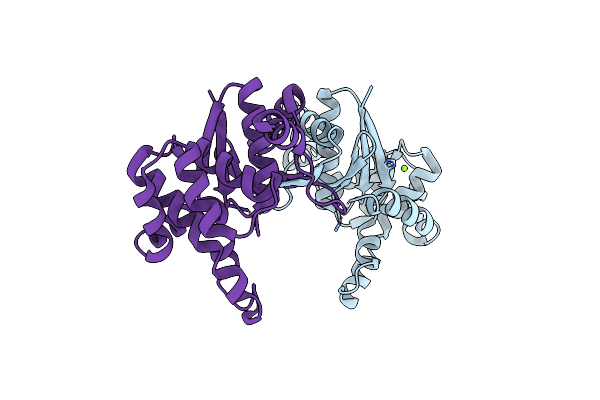
Crystal Structure Of Glucosyl-3-Phosphoglycerate Synthase From Mycobacterium Tuberculosis In Complex With Uridine-Diphosphate (Udp) - Gpgs*Udp
Organism: Mycobacterium bovis af2122/97
Method: X-RAY DIFFRACTION Resolution:2.59 Å Release Date: 2017-05-24 Classification: TRANSFERASE Ligands: UDP |
 |
Organism: Escherichia coli k-12
Method: X-RAY DIFFRACTION Resolution:3.09 Å Release Date: 2017-03-01 Classification: TRANSFERASE |
 |
Crystal Structure Of Glucosyl-3-Phosphoglycerate Synthase From Mycobacterium Tuberculosis - Apo Form
Organism: Mycobacterium tuberculosis h37ra
Method: X-RAY DIFFRACTION Release Date: 2016-12-21 Classification: TRANSFERASE Ligands: GOL |
 |
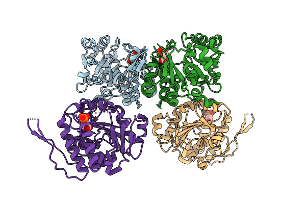
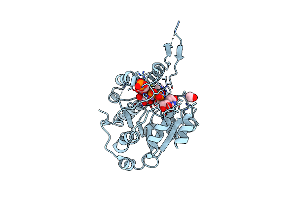
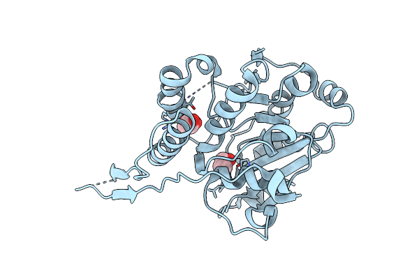
Crystal Structure Of E. Coli Adp-Glucose Pyrophosphorylase (Agpase) In Complex With A Positive Allosteric Regulator Beta-Fructose-1,6-Diphosphate (Fbp) - Agpase*Fbp
Organism: Escherichia coli k-12
Method: X-RAY DIFFRACTION Resolution:3.04 Å Release Date: 2016-09-07 Classification: TRANSFERASE Ligands: SO4, FBP |
 |
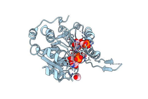
Crystal Structure Of E. Coli Adp-Glucose Pyrophosphorylase (Agpase) In Complex With A Negative Allosteric Regulator Adenosine Monophosphate (Amp) - Agpase*Amp
Organism: Escherichia coli k-12
Method: X-RAY DIFFRACTION Resolution:2.67 Å Release Date: 2016-09-07 Classification: TRANSFERASE Ligands: AMP, PO4 |
 |
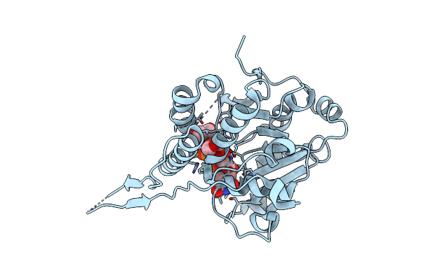
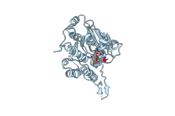
Crystal Structure Of Rv2466C And Oxidoreductase From Mycobacterium Tuberculosis In Its Reduced State
Organism: Mycobacterium tuberculosis (h37rv)
Method: X-RAY DIFFRACTION Resolution:1.51 Å Release Date: 2015-11-18 Classification: OXIDOREDUCTASE Ligands: MG |
 |
Crystal Structure Of Glucosyl-3-Phosphoglycerate Synthase From Mycobacterium Tuberculosis In Complex With Mn2+, Uridine-Diphosphate-Glucose (Udp-Glc) And Phosphoglyceric Acid (Pga) - Gpgs Mn2+ Udp-Glc Pga-1
Organism: Mycobacterium tuberculosis
Method: X-RAY DIFFRACTION Resolution:2.35 Å Release Date: 2015-07-15 Classification: TRANSFERASE Ligands: MN, 3PG, UPG, EDO |
 |
Organism: Mycobacterium tuberculosis (strain atcc 25618 / h37rv)
Method: X-RAY DIFFRACTION Resolution:2.27 Å Release Date: 2015-07-15 Classification: TRANSFERASE Ligands: MN, CL, 3PG, UPG, EDO |
 |
Crystal Structure Of Glucosyl-3-Phosphoglycerate Synthase From Mycobacterium Tuberculosis In Complex With Mn2+, Uridine-Diphosphate-Glucose (Udp-Glc) And 3-(Phosphonooxy)Propanoic Acid (Ppa) - Gpgs Mn2+ Udp-Glc Ppa
Organism: Mycobacterium tuberculosis (strain atcc 25618 / h37rv)
Method: X-RAY DIFFRACTION Resolution:3.23 Å Release Date: 2015-07-15 Classification: TRANSFERASE Ligands: MN, 48X, UPG, SO4, EDO |
 |
Crystal Structure Of Glucosyl-3-Phosphoglycerate Synthase From Mycobacterium Tuberculosis In Complex With Mn2+, Uridine-Diphosphate-Glucose (Udp-Glc) And Glycerol 3-Phosphate (G3P) - Gpgs Mn2+ Udp-Glc G3P
Organism: Mycobacterium tuberculosis (strain atcc 25618 / h37rv)
Method: X-RAY DIFFRACTION Resolution:2.59 Å Release Date: 2015-07-15 Classification: TRANSFERASE Ligands: MN, G3P, UPG |